Alzheimer's Disease Research: Pricella® Cell Lines: Your Trusted Partner for In Vitro Models
Nov 18,2025
In the field of neuroscience, Alzheimer's disease (AD) has long been a focal point of cutting-edge research. As a neurodegenerative disorder characterized by progressive cognitive decline, the majority of currently available drugs can only alleviate symptoms or slow disease progression, and cannot fundamentally reverse it. Therefore, the development of more effective and targeted therapeutic strategies remains an urgent challenge.
In-depth studies of AD pathogenesis rely heavily on in vitro cell models[1-3]. Among these, clonal cell lines have become widely used research tools due to their advantages, including ease of access, rapid growth, straightforward observation, quick isolation, and the ability to provide large quantities of uniform cells through continuous subculturing. Pricella® is dedicated to the research and development of high-quality, stable, and reliable cell lines, and provides comprehensive technical support for cell culture. In this issue of Cell Culture Academy, we will introduce several commonly used cell lines for AD research and highlight their key characteristics.
I. SH-SY5Y
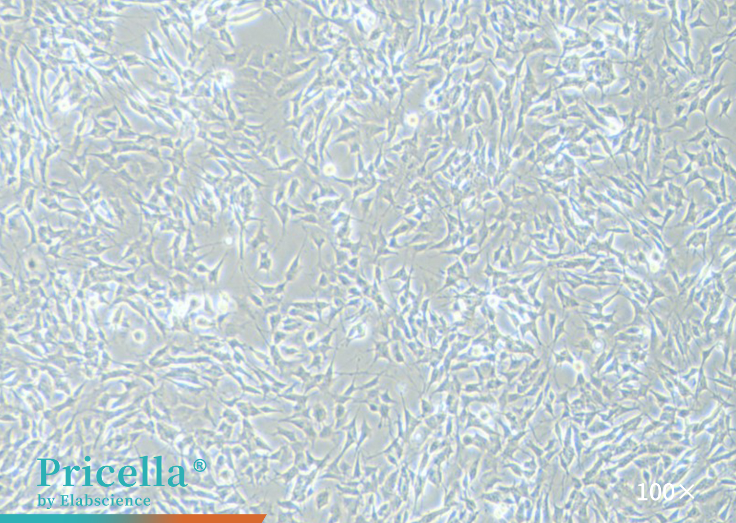
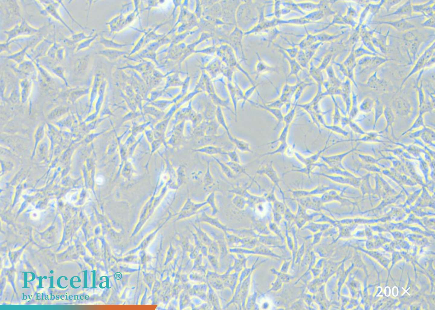
Figure 1. Microscopic Observation of SH-SY5Y
1.Background
SH-SY5Y cells are a triply cloned subline derived from the human neuroblastoma cell line SK-N-SH. This lineage was originally established in 1970 from a metastatic bone tumor in a 4-year-old patient.
2.Applications
Undifferentiated SH-SY5Y cells exhibit continuous proliferation and express immature neuronal markers. Upon induction with agents such as retinoic acid (RA) or brain-derived neurotrophic factor (BDNF), they can differentiate into a neuron-like phenotype. Following differentiation, the cells reproduce key features of cholinergic neurons associated with AD and are widely used as in vitro models in neurodegenerative disease research[4].
3.Culture System
SH-SY5Y [SHSY-5Y] Cell Complete Medium.
4.Key Culture Points
A. SH-SY5Y cells are temperature-sensitive and may detach after shipment at room temperature.
B. Avoid over-digestion, as prolonged enzymatic exposure can compromise cell viability. Repeated or continuous digestion is not recommended, as it may result in suboptimal cell morphology or growth.
C. These cells proliferate relatively slowly; therefore, maintain an adequate seeding density during subculturing to ensure healthy growth.
5.Representative Study
In an Aβ1-42 induced SH-SY5Y cell model, the B14 derivative compound was shown to preserve mitochondrial function and quantity, regulate intracellular reactive oxygen species (ROS) and Ca2+ homeostasis, inhibit Aβ1-42 aggregation, and consequently reduce neuronal apoptosis and delay AD progression [4].
Ⅱ. Neuro-2a
.jpg)
.jpg)
Figure 2. Microscopic Observation of Neuro-2a
1.Background
Neuro-2a cells were established by R.J. Klebe and F. H. Ruddle from a spontaneous neuroblastoma of an A-strain albino mouse.
2.Applications
Neuro-2a cells express high levels of tubulin, which contributes to axoplasmic transport and regulates intracellular trafficking in neurons. When induced by specific stimuli(e.g., colchicine), Neuro-2a cells serve as an in vitro model for studying abnormal Tau or tubulin metabolism and related signaling pathway changes, mimicking the early neurodegenerative state of AD neurons.
3.Culture System
MEM, with NEAA + 10% FBS + 1% Penicillin-Streptomycin Solution.
4.Key Culture Points
A. Cell morphology is heterogeneous, consisting of a mixed population of round and polygonal cells with neurite-like extensions.
B. Cells proliferate slowly; at a 1:2 split ratio, the subculture interval is typically 3-4 days. Maintain adequate cell density to prevent growth inhibition.
C. Cells tend to form clusters or multilayer aggregates and may not uniformly cover the culture surface. Actual cell density is often higher than it appears under the microscope.
5. Representative Study
In a colchicine (OA)-induced Neuro-2a cell model, Lars2 was shown to regulate glycogen synthase kinase 3β (GSK3β) expression via the PI3K-AKT pathway, modulating Tau phosphorylation and promoting neurodegenerative changes[5].
Ⅲ.BV-2
.jpg)
.jpg)
Figure 3. Microscopic Observation of BV-2
1.Background
BV-2 cells were established by E. Blasi in 1990. This immortalized cell line was generated via retrovirus-mediated transfection of v-raf/v-myc into mouse microglial cells and retains key morphological, phenotypic, and functional characteristics of primary microglia.
2.Applications
Microglial cells are the primary immune cells in the brain, playing a central role in defending against injury and infectious agents. BV-2 cells are often co-cultured with neuronal cells(e.g., HT22) to construct neuroinflammation-neuronal injury models that mimic the in vivo environment of AD[6].
3.Culture System
DMEM (High glucose) + 10% FBS + 1% Penicillin-Streptomycin Solution.
4.Key Culture Points
A.BV-2 cells exist in a semi-adherent, semi-suspended state, and the ratio of suspended to adherent cells is variable.
B. During subculturing, collect and process suspended and adherent cells separately. Adherent cells should be digested with 0.25% trypsin with EDTA.
C. Maintain sufficient cell density during subculturing. Cells cultured at low density tend to develop filamentous morphologies with extended processes.
5. Representative Study
In a co-culture model of BV-2 and HT22 cells, stimulation with Aβ25-35 and upregulation of tRFAla-AGC-3-M8 led to increased EphA7 expression in both microglial and neuronal cells, accompanied by elevated phosphorylation of ERK1/2 and p70S6K. Inhibition of EphA7, combined with increased tRFAla-AGC-3-M8 expression, suppressed the ERK1/2-p70S6K signaling pathway in BV-2 and HT22 cells, mitigating neuronal damage, reducing excessive Tau phosphorylation in HT22 cells, and decreasing Aβ25-35 induced M1-type polarization of BV-2 cells[6].
IV. HT-22
.jpg)
.jpg)
Figure 4. Microscopic Observation of HT-22
1.Background
HT-22 cells are a subclone derived from the mouse hippocampal neuronal cell line HT4 and play a critical role in neuropharmacological research.
2.Applications
HT-22 cells exhibit a neuronal phenotype and are highly sensitive to glutamate. Leveraging this property, the glutamate-induced HT-22 neurotoxicity model has become a widely used in vitro model for studying neurotoxicity associated with neurodegenerative diseases, including AD.
3.Culture System
DMEM (High glucose) + 10% FBS+ 1% Penicillin-Streptomycin Solution.
4.Key Culture Points
HT-22 cells are adherent and proliferate relatively quickly.
During cell digestion, closely monitor the digestion state. Avoid over-digestion, as it may compromise cell viability.
5.Representative Study
3-Hydroxyquininedione, a dual inhibitor of ferroptosis and monoamine oxidase B, was evaluated for its anti-ferroptosis activity in HT-22 cells. Its ability to reduce amyloid precursor protein and hyperphosphorylated tau associated with AD was further validated in vivo[7].
Behind every research breakthrough, from in-depth analysis of AD pathological mechanisms to preliminary validation of candidate drugs, lies a stable and reliable in vitro cell model. With strict cell line quality control and professional pre- and post-sales support, Pricella® addresses common challenges such as inconsistent cell performance and experimental variability. Our team also provides personalized culture guidance, offering robust technical support for every stage of AD research.
In addition to its core cell lines optimized for AD research, Pricella® provides well-established neural models such as SK-N-SH and PC-12 to support studies of various neurodegenerative disorders. Dedicated to advancing neuroscience, Pricella® delivers high-performance, quality-controlled cell lines that help researchers overcome experimental barriers, accelerate clinical translation, and fuel reliable in vitro innovation for understanding and treating diseases like Alzheimer’s and Parkinson’s.
References
1.Rumiana TenchoV, et al. Alzheimer’s Disease: Exploring the landscape of cognitive decline. ACS Chemical Neuroscience. 2024, 15, 3800-3827.
2.Targett IL, et al. Differentiation of SH-SY5Y neuroblastoma cells using retinoic acid and BDNF: a model for neuronal and synaptic differentiation in neurodegeneration. In Vitro Cellular & Developmental Biology-Animal. 2024 October;60(9):1058–1067.
3.Anchalee Prasansuklab, et al. Transcriptomic analysis of glutamate-induced HT22 neurotoxicity as a model for screening anti-Alzheimer’s drugs. Scientific Reports. 2023 May 4;13(1): 7225.
4.Dongsheng Ji, et al. Efficient strategy for alleviating neuronal apoptosis and oxidative stress damage of Alzheimer's disease through dual targeting BCL-2 gene promoter i-motif and β-amyloid. Redox Biology, 2025; 82: 103600.
5.Wenqi Qian, et al. Regulating Lars2 in mitochondria: A potential Alzheimer's therapy by inhibiting tau phosphorylation. Neurotherapeutics. 2024 April 4; 21 (4): e00353.
6.Zihao Deng, et al. tRFAla-AGC-3-M8 attenuates neuroinflammation and neuronal damage in Alzheimer's disease via the EphA7-ERK1/2-p70S6K signaling pathway. Alzheimer's Research & Therapy. 2025; 17 (1): 104.
7.Yangjing Lv, et al. 3-Hydroxyquinolin-2-Ones act as dual inhibitors of ferroptosis and monoamine oxidase B: reducing alzheimer’s disease-related amyloid precursor protein and hyperphosphorylated tau In vivo. Journal of Medicinal Chemistry. 2025; 68(13): 13661-13682.
Next: Transduction Protocol for RAW 264.7 Cells: A Complete, Ready-to-use Workflow

