2D vs 3D Cell Culture: Demystifying Core Tumor Models for Modern Cancer Research
May 19,2026
For decades, cancer research has relied on traditional 2D cell culture in Petri dishes, where cells grow as flat monolayers. While this approach has contributed significantly to our understanding of cancer biology, its limitations in mimicking the complex tumor microenvironment have become increasingly apparent. The emergence of 3D culture systems represents a paradigm shift, offering more physiologically relevant models that better recapitulate in vivo tumor conditions[1].
This article explores the fundamental differences between 2D and 3D cell culture methodologies, highlighting why three-dimensional models are revolutionizing cancer research. From tumor organoids to tumor spheroids, these advanced 3D cancer models are bridging the gap between traditional cell culture and clinical reality.
Table of Contents
1.Why 3D Culture is Revolutionizing Cancer Research
2.Key Differences Between 2D Monolayer and 3D Organoid Cultures
3.Comparing Tumor Organoids, Spheroids, and 3D Cancer Models
4.Stem Cell Source Selection for Organoid Derivation
5.Challenges, Advances, and Future Directions in 3D Organoid-Based Cancer Research
01 Why 3D Culture is Revolutionizing Cancer Research
Conventional 2D monolayer cultures fail to replicate the complex three-dimensional architecture, cell-cell interactions, and microenvironmental cues essential for tumor development. This artificial environment often leads to misleading drug screening results and poor clinical translation.
3D culture systems address these limitations by recreating the tumor microenvironment in vitro. Cells interact in all three dimensions, mimicking spatial organization found in actual tumors[2]. This results in more physiologically relevant data with better clinical predictive value.
Key advantages of 3D cell culture include:
Enhanced Drug Response Prediction: 3D models show greater correlation with clinical outcomes.
Tumor Heterogeneity Preservation: Maintain diverse cell populations found in real tumors[3].
Microenvironment Recreation: Incorporate stromal cells, immune cells, and extracellular matrix.
Personalized Medicine Applications: Patient-derived organoids enable tailored treatment strategies.
The transition from 2D to 3D represents a fundamental shift toward more translational cancer research.
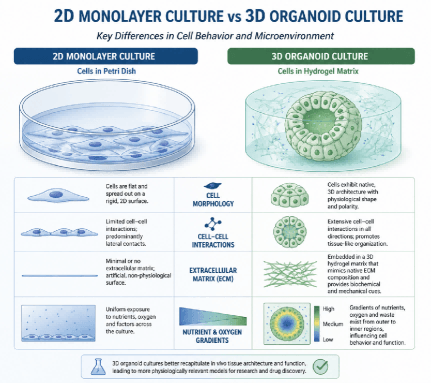
Fig. 1 Comparison of 2D vs 3D cell culture systems.
02 Key Differences Between 2D Monolayer and 3D Organoid Cultures
Understanding the distinctions between traditional 2D cultures and modern 3D cell culture approaches is essential for appreciating their respective applications.
Table 1. Structural and Functional Comparisons
| Feature | 2D Monolayer Culture | 3D Organoid Culture |
| Architecture | Flat, single layer | Complex 3D structure |
| Cell-Cell Interactions | Limited, lateral | Multi-directional, physiological |
| Microenvironment | Artificial, homogeneous | Natural, heterogeneous |
| Polarity | Often lost | Maintained |
| Gene Expression | Altered | More representative |
| Drug Penetration | Uniform exposure | Gradient formation |
| Physiological Relevance | Limited | High |
The table illustrates why organoid culture systems are gaining traction. While 2D cultures remain valuable for high-throughput screening, 3D culture systems offer superior physiological relevance for studying complex tumor behaviors.
Implementing 3D cell culture requires specialized techniques including scaffold selection (hydrogels, synthetic matrices, or scaffold-free systems), oxygen gradient management, and advanced imaging approaches. Despite these challenges, the benefits of accurate tumor modeling justify the investment.
03 Comparing Tumor Organoids, Spheroids, and 3D Cancer Models
The shift from traditional 2D monolayers to 3D in vitro models has been a game-changer in oncology drug development. While 2D cultures have served as the historical standard, they often fail to recapitulate the complex architecture and microenvironment of real tumors. Today, researchers have a spectrum of 3D options. Understanding the nuances between tumor spheroids, tumor organoids, and the broader category of advanced 3D cancer models is crucial for selecting the right tool for your specific biological question.
3.1 Tumor Spheroids: The High-Throughput Workhorse
Tumor spheroids are essentially spherical aggregates of cells. They are typically formed through self-assembly, often driven by cell-cell adhesion forces rather than intrinsic self-organization.
● Structure & Composition: Spheroids are generally simpler structures. They can be derived from cancer cell lines (monocultures) or primary cells. While they successfully mimic the physical gradients of a tumor—such as a proliferating outer layer, a quiescent middle zone, and a necrotic or hypoxic core—they often lack the diverse cellular identity of a real tumor.
● Key Advantages: Their primary strength lies in simplicity and scalability. Spheroids are relatively easy to generate, cost-effective, and highly amenable to high-throughput screening (HTS). They are excellent for initial drug penetration studies and basic toxicity assays.
● Limitations: Because they are often formed from cell lines selected for rapid proliferation, they may not fully capture the genetic and phenotypic heterogeneity of the patient's original tumor.
3.2 Tumor Organoids: The "Patient in a Dish"
Tumor organoids represent a more sophisticated leap in 3D modeling. They are miniaturized and simplified versions of an organ or tumor, produced in vitro in 3D environments.
● Structure & Composition: Unlike spheroids, organoids are driven by the self-organization capabilities of stem cells (either adult tissue stem cells or pluripotent stem cells). They recapitulate the key functional and structural aspects of the tissue of origin, including complex architecture and multiple specialized cell lineages.
● Key Advantages: Organoids offer high clinical predictivity. They faithfully retain the genetic mutations and histological characteristics of the parent tumor, making them powerful tools for precision medicine, biomarker discovery, and modeling patient-specific drug responses over the long term.
● Limitations: They can be more expensive and technically demanding to culture than spheroids. Additionally, standard organoid cultures sometimes lack the full tumor microenvironment (TME), such as immune cells and vasculature, unless specifically engineered via co-culture.
3.3 Advanced 3D Cancer Models: Bridging the Gap
"3D Cancer Models" is the umbrella term, but it also encompasses advanced, engineered systems designed to address specific limitations of spheroids and organoids. These include Patient-Derived Tumor Fragments (PDTFs), Organ-on-a-Chip systems, and 3D Bioprinted models.
● Structure & Composition: These models prioritize the preservation or reconstruction of the Tumor Microenvironment (TME). For instance, tumor fragments are cultured directly without enzymatic dissociation, preserving the native immune cells and stromal architecture. Organ-on-a-chip platforms use microfluidics to introduce dynamic flow and mechanical stress.
● Key Advantages: These are the gold standard for studying complex interactions, such as immunotherapy responses (e.g., CAR-T cell infiltration) or the physical barrier of fibrosis. They provide the most "physiologically relevant" snapshot of the tumor ecosystem.
● Limitations: They often suffer from low throughput, short culture lifespans (in the case of fragments), and high technical complexity, making them better suited for mechanistic studies rather than large-scale drug screening.
If your goal is rapid, large-scale drug screening, spheroids remain a robust choice. If you need to model patient-specific genetics and long-term tumor biology, organoids are superior. However, if your research hinges on the complex interplay between the tumor and its microenvironment (such as immune response), advanced 3D models like tumor fragments or organ-on-a-chip systems are the most appropriate tools.
04 Stem Cell Source Selection for Organoid Derivation
The success of organoid culture depends critically on stem cell source selection.
4.1 Patient-Derived Organoids: Clinical Relevance
Patient-derived organoids represent the gold standard for personalized medicine. Derived directly from patient tumor biopsies, these models preserve genetic and phenotypic characteristics of the original tumor. They are invaluable for predicting individual patient responses, studying tumor heterogeneity, and developing personalized treatment strategies[7].
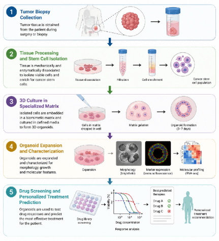
Fig. 2 Patient-derived organoid workflow.
4.2 Stem Cell Derived Organoids: Experimental Control
Stem cell derived organoids can be generated from pluripotent or adult stem cells[4]. These approaches offer greater experimental control and enable the study of tumor development from defined starting populations. They are particularly useful for modeling early tumorigenesis, studying genetic mutations, and creating standardized model systems.
Patient-derived organoids offer the highest clinical relevance and preserve tumor heterogeneity but may be challenging to establish[5][6]. Stem Cell Derived Organoids provide greater experimental control and reproducibility but may lack full tumor complexity[8].
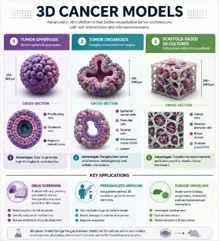
Fig. 3 Types of 3D cancer models.
05 Challenges, Advances, and Future Directions in 3D Organoid-Based Cancer Research
Despite remarkable progress, several challenges remain in 3D cell culture technologies.
5.1 Current Challenges
● Standardization: Variability in protocols affects reproducibility
● Throughput Limitations: More complex than 2D cultures
● Imaging and Analysis: Specialized techniques required
● Cost and Resources: Higher expenses for specialized materials
● Vascularization: Most models lack functional blood vessels
● Immune Component Integration: Incorporating immune cells remains challenging
5.2 Recent Advances
Significant progress includes automation and robotics for high-throughput 3D culture systems, advanced imaging techniques like light-sheet microscopy, microfluidic platforms for better microenvironmental control, standardized protocols for organoid culture, and multi-omics integration with 3D models[9].
5.3 Future Directions
The future of 3D cancer models includes vascularized organoids for studying metastasis, immuno-oncology models with integrated immune cells, multi-organ systems for studying systemic effects, AI and machine learning for predictive modeling, expanded clinical use of patient-derived organoids, and large-scale biobanking initiatives.
Conclusion
The evolution from 2D vs 3D cell culture represents a fundamental shift in cancer research methodology. While traditional monolayer cultures remain valuable for certain applications, 3D culture systems offer unprecedented opportunities to study tumor biology in physiologically relevant contexts.
Tumor organoids, tumor spheroids, and other 3D cancer models are transforming our understanding of cancer progression and therapeutic responses. Whether derived from patient samples or stem cells, these advanced models bridge the gap between basic research and clinical applications.
As technology advances and challenges are addressed, 3D cell culture will play an increasingly central role in cancer research, accelerating the development of more effective treatments and personalized medicine approaches.
References
[1] Lancaster M A, Knoblich J A. Organogenesis in a dish: Modeling development and disease using organoid technologies[J]. Science, 2014, 345(6194): 1247125.
[2] Driehuis E, Clevers H. Cancer modeling meets human organoid technology[J]. Science, 2019, 365(6450): 236-237.
[3] Sachs N, Clevers H. Organoid cultures for the analysis of cancer phenotypes[J]. Current Opinion in Genetics & Development, 2014, 24: 68-73.
[4] Huang L, Holtzinger A, Jagan I, et al. Ductal pancreatic cancer modeling and drug screening using human pluripotent stem cell- and patient-derived tumor organoids[J]. Nature Medicine, 2015, 21(11): 1364-1371.
[5] Vlachogiannis G, Hedayat S, Vatsiou A, et al. Patient-derived organoids model treatment response of metastatic gastrointestinal cancers[J]. Science, 2018, 359(6378): 920-926.
[6] Weeber F, Ooft S N, Dijkstra K K, et al. Tumor organoids as a pre-clinical cancer model for drug discovery[J]. Cell Chemical Biology, 2017, 24(9): 1092-1100.
[7] Sachs N, de Ligt J, Kopper O, et al. A living biobank of breast cancer organoids captures disease heterogeneity[J]. Cell, 2018, 172(1-2): 373-386.
[8] Clevers H. Modeling development and disease with organoids[J]. Cell, 2016, 165(7): 1586-1597.
[9] Boj S F, Hwang C I, Baker L A, et al. Organoid models of human and mouse ductal pancreatic cancer[J]. Cell, 2015, 160(1-2): 324-338.
Prev: Why Do Angiogenesis Assays Fail? A Guide to Experimental Success
Next: Optimizing in Vitro T Cell Culture: A Step-by-Step Experimental Protocol

