Bridging the In Vitro–In Vivo Gap: 3D Tumor Spheroids for Anticancer Drug Screening
Nov 18,2026
After being delivered through the vasculature, drug molecules must penetrate the extracellular matrix, multiple cell layers, and intercellular spaces to reach target cells deep within a tumor. Each physiological barrier and concentration gradient along this route can influence drug efficacy and toxicity. Conventional two-dimensional (2D) monolayer culture cannot reproduce these spatially organized barriers or adequately recapitulate the three-dimensional architecture, cell–cell interactions, and microenvironmental gradients of solid tumors. This helps explain why many anticancer drug candidates that show promising results in 2D cell-based and animal studies fail in clinical development. Three-dimensional (3D) tumor spheroid models are emerging as valuable tools for bridging the in vitro–in vivo gap and supporting anticancer drug screening.
In this edition of Cell Culture Academy, we explore the basic characteristics of 3D tumor spheroids, their key advantages in drug screening, and the principles, strengths, limitations, and applications of different spheroid formation methods.
1. What Are 3D Tumor Spheroids?
3D tumor spheroids are multicellular, spherical structures formed through the spontaneous or induced aggregation of tumor cells. Unlike flat 2D monolayers, spheroids develop spatial gradients of nutrients, oxygen, metabolic waste, and drug concentrations. They also retain cell–cell and cell–matrix interactions that more closely resemble those found under physiological conditions. As a result, cells within spheroids can exhibit tumor-like phenotypes, including reduced proliferation, hypoxia, resistance to apoptosis, and dormancy. For these reasons, tumor spheroids are often regarded as “mini-tumors in vitro.”
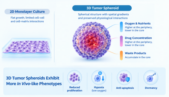
Figure 1. Comparison of 2D monolayer culture and 3D tumor spheroid models
2. Key Advantages of 3D Tumor Spheroids in Drug Screening
3D tumor spheroids support a range of research applications, including high-throughput drug efficacy evaluation, tumor biology studies, tumor microenvironment co-culture, drug sensitivity testing using patient-derived cells, drug toxicity assessment, and evaluation of nanoparticle delivery and penetration. Among these applications, drug screening is one of the most established and commercially relevant.
Compared with conventional 2D culture systems, 3D tumor spheroids offer five major advantages in drug screening:
Recreating In Vivo Spatial and Microenvironmental Gradients
In 2D culture, cells have relatively uniform access to drugs, nutrients, and oxygen. In contrast, as 3D tumor spheroids reach a certain size or are embedded in a matrix, restricted mass transport can generate gradients in oxygenation, metabolites, pH, and drug exposure. This spatial heterogeneity may limit drug penetration into the spheroid core and induce localized cellular tolerance, allowing certain cell subpopulations to survive. By simulating these penetration barriers and microenvironment-dependent differences in drug response at an early stage of drug development, 3D models provide a more physiologically relevant basis for evaluating tissue penetration and overall tumor-killing effects.
More Faithfully Reflecting Tumor Drug Resistance
Compared with cells grown in 2D culture, 3D tumor spheroids often show greater tolerance to a range of chemotherapeutic agents, including cisplatin, paclitaxel, doxorubicin, and 5-fluorouracil. This difference is closely associated with factors such as hypoxia in the spheroid core, reduced cell proliferation, a tendency toward apoptosis resistance, enrichment of stem-like subpopulations, and limited drug penetration into deeper regions of the spheroid. 3D models can therefore better capture tumor cell responses under complex microenvironmental conditions, providing a more physiologically relevant reference for drug sensitivity assessment and candidate screening.
Greater Clinical Relevance
Gene expression profiles, metabolism, and stress-response programs in 3D tumor spheroids more closely resemble those observed in clinical tumors. These models can capture region-specific responses between the outer and inner spheroid layers, as well as reversible, pre-existing drug-tolerant states. This is one reason pharmaceutical companies are increasingly using 3D models in preclinical efficacy studies and investigations of drug resistance mechanisms.
Efficient, Reproducible, and Compatible with High-Throughput Screening
Platforms such as ultra-low attachment (ULA) 384-well plates enable standardized spheroid production with improved batch-to-batch consistency and compatibility with automated liquid handling. They also support long-term monitoring of drug responses and the detection of recurrence from drug-resistant subpopulations. These capabilities are particularly valuable for primary screening of large compound libraries and dose–response modeling.
Reducing Development Time and Costs
Eliminating ineffective or highly drug-resistant candidates at an earlier stage can reduce the need for animal studies and costly clinical trial-and-error. Compared with patient-derived xenograft (PDX) models, cell line-derived 3D spheroids offer a practical balance between physiological relevance, ease of use, and cost-effectiveness, making them useful early-stage models in the drug development pipeline.
3. Comparison of Different 3D Tumor Spheroid Formation Methods
The biological characteristics of 3D tumor spheroids, including compactness, structural uniformity, and drug responses, are influenced by the method used to generate them. Different methods may produce distinct patterns of cell organization, extracellular matrix remodeling, and drug sensitivity. The table below summarizes the principles, advantages, limitations, and typical applications of commonly used spheroid formation methods.
| Formation Method | Principle | Advantages | Limitations | Typical Applications |
| Ultra-Low Attachment (ULA) Plates(Forced-Floating Culture) | Non-adhesive surfaces prevent cell attachment, allowing cells to aggregate spontaneously into spheroids. | Suitable for large-scale production; low cost; simple operation; readily compatible with high-throughput screening in 384-well plates; convenient compound addition and medium exchange. | Some cell lines form loose spheroids with insufficient compactness. | High-throughput drug screening; batch spheroid production. |
| Hanging Drop Method | Surface tension and gravity within droplets drive cell aggregation. | Up to 384 spheroids can be generated in a single run; spheroid size and cell composition can be controlled; no specialized equipment required. | Spheroid formation is difficult to track; compound addition and medium exchange are inconvenient; droplets may evaporate; labor-intensive and difficult to scale up. | Small-scale studies requiring strict control of spheroid size. |
| Spinner Flasks / Bioreactors | Continuous stirring or rotation prevents cell attachment and promotes aggregation. | Suitable for large-scale spheroid production. | Inappropriate rotation speeds may damage or disperse cells; spheroid diameters may vary; specialized equipment is required; real-time observation is difficult. | Large-volume production and scale-up. |
| Microfluidic Chips | A 3D environment is created and perfused within microchannels. | Can simulate vascularization and perfusion; supports long-term culture; precisely controlled conditions; suitable for high-throughput applications. | Requires specialized equipment and training; polydimethylsiloxane (PDMS) may swell, potentially affecting flow rates. | Tumor microenvironment studies, vascularization, and dynamic perfusion. |
| Matrix Embedding(Matrigel / Hydrogels) | Cells are embedded and grown within a matrix. | Preserves cell–matrix interactions and more closely resembles physiological conditions. | Restricted diffusion may increase the risk of cell death; matrix handling is complex. | Invasion and migration assays; cell–matrix interaction studies. |
| Magnetic Levitation / Magnetic 3D Bioprinting | Cells are labeled with magnetic nanoparticles and assembled into floating spheroids. | Facilitates the formation of heterogeneous spheroids; rapid spheroid formation. | Requires preparation of magnetic particles; scalability is limited. | Heterogeneity studies and co-culture models. |
| Micropatterned Culture Plates | Patterned surfaces guide cells to form spheroids at defined locations. | Scalable; low well-to-well and plate-to-plate variability. | Spheroid diameters may be uneven; bubbles may form; pipetting may damage the patterns. | Standardized high-throughput applications. |
Why Does Procell Choose the ULA Plate Method?
ULA plates offer several advantages, including ease of use, controllable costs, and suitability for high-throughput screening. However, spheroids formed by some cell lines may be loose or insufficiently uniform. To address these challenges, Procell has developed dedicated culture reagents and conducted systematic screening and optimization of culture conditions, with a focus on improving spheroid compactness, uniformity, and cell viability. This approach enhances the quality of spheroid model generation while retaining the operational simplicity and throughput advantages of the ULA method.
3D tumor spheroids provide a more physiologically relevant in vitro model for anticancer drug screening, enabling a more comprehensive assessment of candidate compounds in terms of drug penetration, tumor resistance, and pharmacological response. Each formation method has its own characteristics, and researchers should select an appropriate strategy based on the cell type, research objectives, and throughput requirements. As 3D culture technologies and evaluation systems continue to advance, tumor spheroid models are expected to provide increasingly valuable evidence for drug development and tumor biology research.
Prev: Difficulty Obtaining Human Neurons? A Complete Guide to NGN2-Induced Neuronal Differentiation