HBSS vs PBS vs DPBS and Which Buffer Is Right for Your Cell Experiment
Oct 12,2026
PBS, DPBS and HBSS are the most commonly used balanced salt solutions in cell biology. PBS is ideal for routine cell washing and dilution. DPBS, which can be formulated with or without calcium and magnesium, is suitable for various cell processing procedures. Containing glucose and bicarbonate, HBSS effectively maintains cell viability for short periods outside the incubator. So how should we choose the right balanced salt solution for cell experiments?
Table of Contents
1. HBSS vs. PBS vs. DPBS vs. saline: what’s the actual difference?
2. Why you can’t always swap one salt solution for another
3. Why calcium and magnesium can change your results
4. Immune cell activation sensitivity to buffer ionic composition
5. How to choose the right salt solution for your experiment
01 HBSS vs. PBS vs. DPBS vs. saline: what’s the actual difference?
In cell culture and translational life science research, selecting an appropriate cell culture buffer is one of the most fundamental yet frequently underestimated steps for generating consistent, reproducible experimental data. Most laboratories routinely employ phosphate buffered saline, HBSS buffer, PBS buffer, or DPBS buffer for routine washing, cell passaging, and sample preparation without a thorough understanding of their compositional and functional differences. These balanced salt solutions are not interchangeable; their distinct ionic profiles directly dictate cell viability, physiological homeostasis, and the reliability of final experimental outcomes[1].
To systematically clarify their functional disparities, it is critical to first define each buffer and compare their core formulations, starting with the simplest option: normal saline. Composed exclusively of 0.9% sodium chloride in ultrapure water, normal saline contains no buffering agents, trace minerals, or energy substrates. While it maintains physiological isotonicity, it lacks the pH stabilization capacity and complex ionic balance required for live cell manipulation, rendering it unsuitable for nearly all long-term cell culture applications.
PBS buffer, fully named phosphate buffered saline, serves as the most widely used general-purpose buffer in biological laboratories. Standard PBS consists of sodium chloride, potassium chloride, and a dual phosphate buffering system comprising sodium dihydrogen phosphate and disodium hydrogen phosphate. Formulated to maintain a stable physiological pH of 7.4 and isotonic pressure, PBS is optimized for brief cell rinsing, reagent dilution, and fixed tissue sample processing. Traditional PBS baseline formulations exclude calcium and magnesium ions, a feature that simplifies long-term storage but limits its compatibility with native live cell physiological microenvironments.
DPBS buffer, or Dulbecco’s Phosphate Buffered Saline, is a refined, cell-culture-specific variant of conventional PBS first developed for mammalian cell culture and viral research[2]. While retaining PBS’s core phosphate buffering mechanism, DPBS features optimized ion concentrations and minimized trace contaminants to meet strict mammalian cell culture standards. A defining distinction in HBSS vs DPBS lies in formulation versatility. Commercially available DPBS is provided in two standardized formats: with or without calcium and magnesium. Cation-free DPBS facilitates efficient cell dissociation and prevents cellular aggregation, whereas cation-supplemented DPBS closely mimics in vivo ionic conditions for physiological cell maintenance.
HBSS buffer, or Hanks’ Balanced Salt Solution, is a physiologically complete buffer with a far more sophisticated composition than standard PBS and DPBS[3]. The comprehensive HBSS composition incorporates sodium chloride, potassium chloride, calcium chloride, magnesium sulfate, sodium bicarbonate, glucose, and phosphate buffer components. Unlike PBS and DPBS, most standard HBSS formulations inherently contain physiological concentrations of calcium and magnesium, commonly referred to as HBSS with calcium and magnesium. This biologically relevant formula closely replicates human interstitial fluid, supporting transient cellular metabolism and membrane stability. The primary discrepancies in HBSS vs PBS stem from HBSS’s unique inclusion of divalent cations, glucose, and bicarbonate, which are essential for sustaining temporary live cell physiological activity[1].
Table 1. Comparison of commonly used balanced salt solutions
| Buffer Solution | Key Components | Contains Ca²⁺/Mg²⁺ | Glucose & Bicarbonate | Primary Application Scenarios |
| Normal Saline | Sodium chloride only | No | No | Short-term tissue rinsing, non-cell biological experiments |
| Standard PBS | NaCl, KCl, phosphate buffer | No | No | Fixed sample washing, reagent dilution, general rinsing |
| DPBS | Optimized PBS formula, low impurities | Optional | No | Cell passaging, live cell washing, flow cytometry sample preparation |
| HBSS | Complete salt system + glucose + bicarbonate | Yes (standard formulation) | Yes | Temporary cell incubation, tissue dissection, physiological simulation |
02 Why you can’t always swap one salt solution for another
A pervasive misconception in cell culture practice is that all isotonic, pH-calibrated salt solutions deliver equivalent experimental performance. Many researchers freely substitute PBS for DPBS or HBSS during routine workflows, assuming negligible impacts on experimental data. In reality, arbitrary interchange of cell culture buffer solutions is a major contributor to inconsistent cell morphology, compromised cell viability, and poor experimental reproducibility across research laboratories.
Metabolic incompatibility represents the primary barrier to unrestricted buffer substitution. HBSS contains glucose and sodium bicarbonate, which provide transient energy supply and synergistic pH buffering for live cells. Cells maintained or rinsed in HBSS can preserve basal metabolic function and membrane integrity for several hours. In contrast, both PBS buffer and DPBS buffer lack glucose and bicarbonate. Prolonged exposure of live cells to these metabolically inert buffers disrupts intracellular energy homeostasis, leading to suppressed cellular activity, cytoplasmic shrinkage, and eventual apoptosis, particularly in sensitive primary cells and suspension cell populations.
Variable ionic tolerance across cell types creates a second critical limitation for buffer substitution. Immortalized adherent cell lines such as HEK293 and HeLa demonstrate robust tolerance to minor buffer discrepancies and remain morphologically stable after short-term exposure to mismatched buffers. This high tolerance often fosters improper substitution habits among researchers. However, primary cells, stem cells, and immune cells exhibit extreme sensitivity to compositional changes in extracellular buffers. Replacing cation-containing HBSS with standard cation-free PBS disrupts membrane adhesion complexes, triggers unintended cell detachment, and impairs the functional conformation of cell surface proteins.
Furthermore, these buffers rely on fundamentally distinct pH buffering mechanisms. PBS and DPBS utilize phosphate-only buffering, which maintains stable pH under ambient atmospheric conditions. HBSS adopts a dual bicarbonate-phosphate buffering system that requires 5% CO₂ incubation to sustain physiological pH levels. When HBSS is used in ambient air without CO₂ supplementation, its pH rapidly elevates, inducing alkaline stress and cellular damage. Conversely, utilizing PBS for long-term live cell assays fails to recapitulate the physiological bicarbonate buffering environment present in vivo, resulting in distorted cellular responses[1]. These inherent functional differences make indiscriminate buffer substitution a significant threat to experimental standardization and reproducibility.
03 Why calcium and magnesium can change your results
Divalent calcium and magnesium ions are not inert additives in balanced salt solutions; they are core modulators of cellular physiological behavior. Their presence or absence constitutes the most impactful variable in HBSS vs PBS and HBSS vs DPBS comparisons. These two cations regulate cell membrane stability, intercellular adhesion, protein conformational integrity, and intracellular signal transduction, ultimately determining the accuracy and consistency of cell-based experimental results.
Calcium ions sustain the structural integrity of cell membrane lipid bilayers and intercellular tight junctions. For adherent cells, extracellular calcium supports the proper folding and activation of integrin proteins, ensuring stable cell attachment to culture substrates. Magnesium ions serve as essential cofactors for the majority of intracellular kinases and metabolic enzymes, regulating cellular metabolism and cytoskeleton organization. HBSS with calcium and magnesium preserves these physiological cation levels, generating an extracellular environment consistent with in vivo interstitial fluid. This formulation effectively maintains native cell morphology and physiological activity during tissue dissection, primary cell isolation, and short-term cellular incubation.
In cell dissociation and passaging workflows, calcium and magnesium act as interfering factors. Divalent cations strengthen cell-cell and cell-matrix adhesion, inhibiting trypsin-mediated breakdown of adhesion proteins. This interference results in incomplete cell dissociation, cellular aggregation, and reduced cell recovery yields. For this reason, calcium- and magnesium-free DPBS is the gold standard for routine cell passaging and single-cell suspension preparation. Using cation-containing standard HBSS for dissociation severely compromises experimental efficiency and introduces variability in cell counting and downstream analysis.
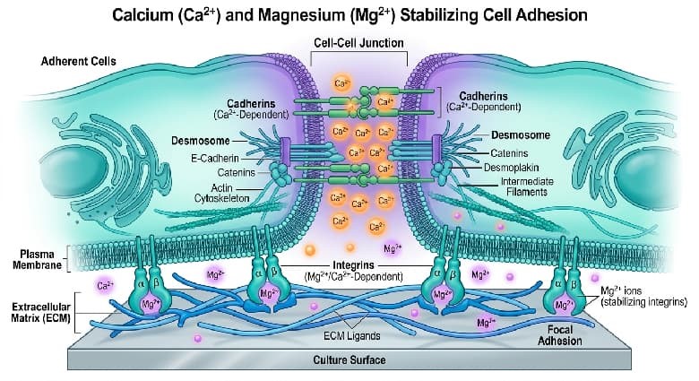
Fig. 1 Calcium and magnesium impact on cell adhesion.
Cation composition also profoundly influences biomolecular activity in experimental systems. Most cell surface receptors, antibodies, and metabolic enzymes exhibit cation-dependent functional activity. Antigen-antibody binding in immunostaining assays and receptor-ligand interactions in cell signaling studies are both tightly regulated by extracellular calcium and magnesium. Mismatched buffer cation concentrations alter protein binding affinity, leading to attenuated experimental signals, elevated background noise, and false positive or negative results. Even minor variations in ionic composition can induce statistically significant bias in quantitative cell-based assays[1].
04 Immune cell activation sensitivity to buffer ionic composition
Immune cell functional assays represent the most sensitive application scenario for cell culture buffer selection. Unlike robust immortalized tumor cell lines, primary immune cells including T lymphocytes, B lymphocytes, macrophages, and dendritic cells are highly susceptible to fluctuations in extracellular ionic conditions[4]. Their activation status, cytokine secretion profiles, and proliferative capacity are readily perturbed by suboptimal buffer environments, making deliberate buffer selection a prerequisite for reliable and reproducible immunological data[4].
The ionic strength and cation content of phosphate buffered saline and HBSS directly modulate immune cell membrane potential and receptor functionality. Cell surface pattern recognition receptors and cytokine receptors rely on physiological ionic concentrations to maintain their native three-dimensional conformation. Resuspending immune cells in low-cation PBS disrupts membrane potential homeostasis, triggering low-level non-specific immune activation. This spontaneous baseline activation elevates endogenous cytokine expression, obscuring true experimental responses and compromising assay accuracy.
HBSS buffer supplemented with physiological calcium and magnesium stabilizes immune cells in a quiescent resting state. Its balanced ionic profile eliminates ionic stress-induced false activation, ensuring that measurable cellular responses derive solely from experimental stimuli rather than buffer artifacts. In contrast, standard cation-free PBS buffer fails to stabilize immune cell membrane structure. Prolonged sample processing in PBS induces cellular fatigue, reduces antigen responsiveness, and promotes aberrant apoptosis, leading to systematic underestimation of immune cell activation levels.
DPBS buffer exhibits context-dependent performance in immune cell assays, determined entirely by its cation formulation. Calcium- and magnesium-free DPBS is ideal for rapid washing and single-cell suspension generation for immune cell samples, as it prevents cellular clumping without inducing significant activation during short-duration processing. However, prolonged incubation in cation-free DPBS impairs immune cell viability and functional activity. For immune cell co-culture, cytokine profiling, and other functional assays, complete HBSS with full ionic supplementation is superior for sample preprocessing and temporary sample storage.
Multiple laboratory validation studies confirm that buffer ionic discrepancies can induce 20% to 30% deviation in quantified immune cell activation rates. This magnitude of error far exceeds the acceptable tolerance range for biological experiments, verifying that buffer selection is not a trivial procedural step but a critical factor for controlling experimental background noise and ensuring data integrity[4].
05 How to choose the right salt solution for your experiment
Selecting the optimal balanced salt solution does not require complex theoretical analysis, yet it demands precise alignment with experimental objectives, cell type specificity, and operational duration. By understanding the core functional distinctions between HBSS vs PBS and HBSS vs DPBS, researchers can establish standardized buffer selection criteria to minimize buffer-induced experimental bias and improve data reproducibility.
Select standard PBS buffer exclusively for fixed biological sample processing and basic rinsing workflows. PBS is well-suited for washing fixed cells and tissue sections, diluting non-biological reagents, and routine surface decontamination of culture vessels. It is unsuitable for all live cell incubation and extended live cell processing due to the absence of energy substrates and cytoprotective ions.
Select DPBS buffer for routine live cell passaging and flow cytometry sample preparation. Calcium- and magnesium-free DPBS is the optimal choice for cell dissociation and single-cell suspension generation, as it eliminates cation-mediated intercellular adhesion. Cation-supplemented DPBS can be used for short-term post-adhesion cell rinsing, balancing operational practicality and cellular protection. Its high purity and consistent formulation make DPBS the most versatile buffer for routine mammalian cell culture maintenance.
Select HBSS buffer (preferably HBSS with calcium and magnesium) for physiological simulation, tissue dissection, primary cell isolation, and temporary live cell incubation. The comprehensive HBSS composition, including glucose, bicarbonate, and physiological divalent cations, sustains cellular metabolic activity and native morphology for 2 to 4 hours. It serves as the preferred buffer for all workflows requiring prolonged cellular activity, including in vitro tissue incubation, primary cell extraction, and real-time functional cell analysis.
For sensitive cell assays including immune cell activation, stem cell culture, and primary cell functional analysis, prioritize fully supplemented HBSS to prevent non-specific cellular activation and activity loss. For quantitative assays such as ELISA and flow cytometry, maintain consistent buffer types across all sample processing steps to ensure uniform ionic background and eliminate systematic experimental error.
In summary, PBS, DPBS, and HBSS each possess unique functional roles in cell culture workflows. No single buffer is universally superior; instead, each is optimized for distinct experimental scenarios. Standardizing buffer selection and application is a low-cost, high-impact strategy to enhance the reproducibility and accuracy of cell culture experiments.
References:
[1] TRIVEDI M, PATEL S. Comparative Physicochemical Evaluation of Phosphate Buffer Saline and Hanks Balanced Salt Medium[J]. Journal of Bioengineering & Biomedical Science, 2015, 5(2): 1-6.
[2] DULBECCO R, VOGT M. Plaque formation and isolation of pure lines with poliomyelitis viruses[J]. Journal of Experimental Medicine, 1954, 99(2): 167-182.
[3] HANKS J H, WALLACE R E. Relation of oxygen and temperature in the preservation of tissues by refrigeration[J]. Proceedings of the Society for Experimental Biology and Medicine, 1949, 71(1): 196-200.
[4] NG S, LEE J. Ionic composition of culture buffers modulates primary immune cell activation and cytokine secretion[J]. Journal of Immunological Methods, 2022, 506: 113289.