Difficulty Obtaining Human Neurons? A Complete Guide to NGN2-Induced Neuronal Differentiation
Nov 04,2026
Neurons are indispensable in vitro models for studying human nervous system development, investigating the mechanisms underlying neurodegenerative diseases, and evaluating drug effects. However, the use of primary human neurons has long been constrained by challenges in obtaining brain tissue, stringent ethical review, limited donor samples, and substantial donor-to-donor variability, all of which can compromise experimental consistency and reproducibility. Although neurons of animal origin are easier to obtain, species differences make it difficult to recapitulate the distinctive physiological and pathological features of the human nervous system, limiting the translation of findings to humans.
Efficiently obtaining human neurons with a stable origin, consistent phenotype, and suitability for in vitro experiments remains a key challenge in neuroscience.
In this edition of Cell Culture Academy, we break down the principles behind NGN2-directed neuronal differentiation, the induction workflow, key culture considerations, and potential research applications.
1. NGN2-Induced Neuronal Differentiation
1.1 Technical Principles
H9 human embryonic stem cells are transduced with a lentiviral vector carrying the Tet-On-inducible NGN2-Puro construct. Neuronal differentiation is then directed using a doxycycline (Dox)-inducible expression system.
In the presence of Dox, the Tet-On system activates NGN2 expression. As a key transcription factor involved in neurogenesis, NGN2 regulates the expression of genes associated with neuronal differentiation, promoting the transition of cells toward a neuronal fate. Puromycin selection is also used to enrich the population carrying the target construct and eliminate cells that have not successfully integrated it.
1.2 Induction Workflow
Day 1 | Cell Seeding
Prepare a six-well plate coated with Matrigel. Dissociate and count H9 cells carrying the integrated NGN2 construct, then seed at 2.4×10⁵ cells per well.
Days 2–3 | Initiate Induction
Replace the H9 cell culture medium with induction medium containing N-2 and add Dox to a final concentration of 2 μg/mL to initiate NGN2 expression. Replace the medium daily during this stage.
Day 4 | Medium Change and Positive Selection
Switch to Neurobasal medium containing B-27 and continue adding Dox to maintain induction.
Add puromycin to a final concentration of 1 μg/mL to select positive cells. Cells that have not been successfully transduced with TetO-NGN2-Puro will gradually die.
Days 5–8 | Continue Induction
Replace the medium with fresh medium every 2 days and continue adding Dox. Before each medium change, slowly add DPBS along the well wall and gently rinse to remove cell clusters that are about to detach. Aspirate the DPBS before adding fresh medium.
Day 8 | Cell Harvest and Cryopreservation
The cells should show clear signs of neuronal differentiation. Depending on downstream experimental needs, they may be cultured further or cryopreserved.
1.3 Neuronal Morphology and Immunofluorescence Characterization
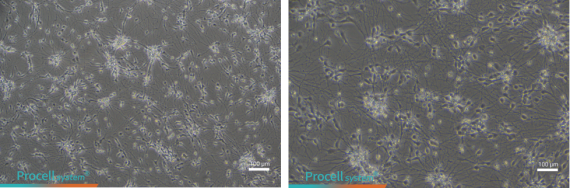
Figure 1. Neuronal morphology on Day 9 of induction in NGN2-overexpressing H9 stem cells
The cell bodies are plump and clustered, while numerous long, slender neurites interconnect to form a dense neuronal network, indicating a mature differentiated morphology and healthy cell state.
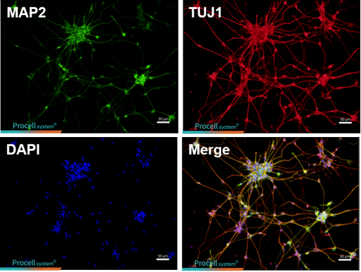
Figure 2. Immunofluorescence characterization on Day 20 of induction in NGN2-overexpressing H9 stem cells
MAP2 (green) is a marker associated with mature neurons and specifically labels neuronal cell bodies and dendrites. TUJ1 (red) is a pan-neuronal marker that labels all neurons, including immature neurons. DAPI (blue) stains cell nuclei. Many cells co-express MAP2 and TUJ1, with abundant neurites forming a neuronal network, supporting the presence of a highly enriched population of mature neurons in a favorable differentiation state.
2. Key Considerations for Culturing NGN2-Induced Neurons
NGN2-induced neurons are sensitive to culture conditions and mechanical stress. They are terminally differentiated cells that do not proliferate and cannot be routinely passaged. Pay particular attention to the following points during thawing and seeding, routine culture, and replating for experiments.
2.1 Thawing and Seeding: Improve Attachment and Survival
Coat culture vessels: Pre-coat culture vessels with 0.1 mg/mL poly-L-lysine (PLL) to improve cell attachment.
Temperature control: Prewarm culture-related reagents to 37°C to minimize the effects of temperature fluctuations on cell condition. Avoid prolonged exposure of cells to room temperature.
Add a cell-protective agent: During thawing and seeding, adding Y-27632 to the medium at a final concentration of 10 μM is recommended to improve post-thaw survival and attachment.
Handle gently: Gently mix the cell suspension and avoid vigorous pipetting to minimize cell damage.
2.2 Routine Culture: Monitor Neurite Growth and Network Formation
Change medium gently: Avoid directing the flow of medium onto the cell layer to reduce neurite breakage and cell detachment.
Monitor cell morphology: Pay close attention to cell body condition, neurite extension, and the formation of networks between cells. If marked cell body shrinkage, broken neurites, or widespread cell detachment occurs, promptly investigate the culture conditions.
Plan experiments carefully: The effective in vitro culture period is approximately 14 days after thawing. Schedule experimental time points according to the research objectives and keep culture duration as consistent as possible across experimental groups.
2.3 Replating: Optimize Dissociation Conditions and Seeding Density
Control dissociation conditions: Use 0.125% trypsin solution, prepared by diluting 0.25% trypsin containing EDTA 1:1 with PBS. Allow the solution to equilibrate to room temperature before use. Dissociate at room temperature for approximately 4–5 minutes, gently rocking the vessel and monitoring cell detachment under a microscope. Do not exceed 5 minutes per dissociation. Stop dissociation immediately when a substantial number of neurons begin to detach.
Avoid mechanical damage: When collecting and resuspending cells, avoid forceful pipetting or scraping to minimize damage to cells and neurites.
Control seeding density: A recommended replating density is 1–1.2 × 10⁵ cells/cm², which can be adjusted to suit the experiment. Culture vessels must be pre-coated with PLL before seeding.
Allow time for recovery: After replating, leave the cells undisturbed for 24–48 hours to allow them to reattach and extend their neurites before proceeding with downstream experiments.
3. Potential Research Applications of NGN2-Induced Neurons
Human cortical excitatory neurons generated by NGN2 induction can be used in a range of neuroscience studies. Based on their cell type and in vitro culture characteristics, potential applications include the following.
3.1 Neuronal Development and Network Research
These neurons can be used to investigate neuronal differentiation and maturation, neurite outgrowth, synapse formation, and neuronal network development, helping characterize the developmental and functional properties of human neurons.
3.2 Mechanistic Studies of Neurological Disorders
With an appropriate disease-associated genetic background or pathological stimulus, these neurons can be used to investigate mechanisms relevant to neurodegenerative diseases such as Alzheimer's disease and amyotrophic lateral sclerosis (ALS). The suitability of a specific model depends on the study design and the characteristics of the cell model itself.
3.3 Neuronal Injury and Stress Research
These neurons can be used to study neuronal responses to oxidative stress, excitotoxicity, and ischemia- or hypoxia-related injury, helping elucidate cellular injury mechanisms and potential intervention targets.
3.4 Drug Screening and Neurotoxicity Assessment
Applications include preliminary in vitro screening of candidate compounds, studies of neuroprotective effects, and neurotoxicity assessment, providing a human cell model for drug development.
Neuronal model-generation technologies continue to evolve, from the use of traditional primary neurons to directed differentiation from stem cells. By regulating the expression of a key transcription factor, NGN2-induced differentiation promotes the transition of cells toward a neuronal fate, offering another model option for research on neuronal development, disease mechanisms, and drug development.
At the same time, experimental outcomes are influenced by differentiation status, cell type, culture conditions, and functional maturity. Understanding the biological characteristics of the model, following key culture practices, and selecting appropriate readouts based on the research objectives are essential for improving reproducibility and obtaining reliable data.