Optogenetics Opens a New Window into Neuroscience
Oct 08,2026
On October 5, 2026, the Nobel Prize in Physiology or Medicine was announced. Peter Hegemann, Georg Nagel, and Karl Deisseroth were recognized for foundational discoveries involving light-gated ion channels and optogenetics.

Figure 1. Recipients of the 2026 Nobel Prize in Physiology or Medicine. Figure from the official Nobel Prize website (www.nobelprize.org)
The origins of this technology can be traced to a single-celled alga that swims toward light.
Researchers identified channelrhodopsin, a light-sensitive protein, in the algal cell membrane. When blue light illuminates the cell, channelrhodopsin is activated and allows ions to cross the membrane, generating an electrical signal.
Researchers later introduced these light-sensitive proteins into mammalian neurons, enabling the neurons to respond to specific wavelengths of light. Light stimulation can then activate or inhibit the activity of targeted neurons on a millisecond timescale.
Light thus became a tool for controlling neural activity, giving rise to optogenetics.
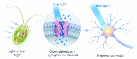
Figure 2. Origins and Basic Principles of Optogenetics
From Neural Stimulation to Precise Control of Neurons
Before optogenetics emerged, neuroscience already relied on methods such as electrical stimulation and pharmacological intervention. However, precisely controlling neural activity remained challenging.
Conventional electrical stimulation often cannot selectively target a specific neuronal type, while pharmacological interventions are limited by their time course and cell-type specificity. These limitations make it difficult to answer questions such as which neurons contribute to a particular behavior or what happens when a specific neuronal population is activated.
Optogenetics changed this.
Using genetic targeting strategies, researchers can drive the expression of light-sensitive proteins in specific neurons, effectively giving the target cells a light-controlled switch. Stimulation with light of a selected wavelength can then modulate neuronal activity with high temporal and cell-type specificity.
This approach allows researchers not only to observe neural activity, but also to actively control the activity of selected neurons and investigate how it relates to neural circuits and behavior.
A Range of Research Applications for Optogenetics
Over the past 20 years, optogenetics has become an important tool in neuroscience. Research has expanded from early analyses of neuronal function to the study of neural circuits, animal behavior, and disease mechanisms. Its experimental platforms now span in vitro cell models, live-animal studies, and translational research.
In Vitro Cell Studies
Light-sensitive proteins can be expressed in cultured neurons or other cell models. Combined with light stimulation, electrophysiology, and calcium imaging, this approach can be used to investigate neuronal excitability, signal transmission, and cellular responses, as well as to validate experimental systems.
Live-Animal Studies
By targeting light-sensitive proteins to specific neuronal populations and delivering light through tools such as optical fibers, researchers can modulate neural circuits with precise timing to investigate complex functions such as memory, emotion, movement, and behavior.
Disease Mechanisms and Translational Research
Optogenetics has also been used in basic research on Parkinson’s disease, epilepsy, retinal degenerative diseases, and other conditions. It has helped advance some light-based neuromodulation strategies toward translational research.
Reliable Cell Models Are Essential for In Vitro Optogenetics
From light-sensitive protein expression and light stimulation to downstream functional assays such as electrophysiological recording and calcium imaging, multiple stages of in vitro optogenetics experiments depend on cell models.
Cell viability, purity, morphology, and biological-state consistency can all affect cellular responses to light stimulation and the results of functional assays. Primary neurons are particularly sensitive to culture conditions, and their physiological state and the formation of neuronal networks can influence downstream experiments.
Therefore, in in vitro optogenetics research, experimental design should account not only for the light-sensitive protein, transfection method, and illumination conditions, but also for selecting a cell model suited to the research question and establishing a stable culture system.
Different Neuroscience Questions Require Different Cell Models
Depending on the research objective, cell models with different biological characteristics may be appropriate:
Hippocampal neurons: commonly used to study learning and memory, synaptic plasticity, and related neural circuits.
Cerebellar granule cells: useful for studies of neural development and cerebellum-related functions.
Olfactory bulb neurons: suitable for research on olfactory pathways and related neural functions.
Neuronal cell lines and other cell lines: useful for preliminary validation and optimization of light-sensitive protein expression, light-stimulation conditions, and experimental workflows.
For in vitro neuroscience and optogenetics research, Procell offers a portfolio of cell resources and culture systems, including a range of primary neuronal cells and commonly used cell lines, together with corresponding media and growth supplements.
From the discovery of light-activated ion channels to the precise optical control of selected neurons, optogenetics has moved neuroscience from observing neural activity toward controlling it.
This beam of light does more than switch neurons on or off. It opens new ways to investigate the complex mechanisms underlying memory, emotion, behavior, and neurological disease. As the technology advances, its integration with diverse cell models, imaging, and electrophysiology will continue to deepen our understanding of the brain and make previously difficult neuroscience questions more tractable.
Prev: In Vitro Cell Models: Powerful Tools for Cardiovascular Disease Research