In Vitro Cell Models: Powerful Tools for Cardiovascular Disease Research
Sep 29,2026
September 29 marks the 26th World Heart Day. This year, the World Heart Federation (WHF) continues the “Don’t Miss a Beat” theme, reminding us to pay attention to easily overlooked signs of cardiovascular health.
Many cardiovascular diseases are clinically silent in their early stages and may present without typical symptoms. By the time clear symptoms appear, irreversible damage may already have occurred. Reliable in vitro models are therefore essential for cardiovascular research, helping researchers capture key changes before disease mechanisms become fully apparent.
In vitro research systems use cardiomyocytes, vascular endothelial cells, vascular smooth muscle cells, cardiac fibroblasts, macrophages, and other cell types as important cellular foundations for studying the cardiovascular system. These cells do not experience chest pain or develop true arrhythmias, but they can recapitulate key biological processes of the heart and cardiovascular system in culture, making them valuable models for investigating disease mechanisms and evaluating drugs and therapeutic strategies.
Procell provides a range of cell models and supporting culture reagents for cardiovascular research. In recent years, related products have been used in studies of myocardial injury, myocardial ischemia, angiogenesis, and cardiovascular disease mechanisms. Here, we highlight three literature examples to see how these cell models contribute to cardiovascular research.
Ⅰ. From Chemotherapy-Induced Cardiac Injury to Mitochondrial Damage Mechanisms
Doxorubicin (Dox) is a widely used chemotherapeutic agent, but its cardiotoxicity remains an important concern in cancer treatment. Researchers established an in vitro model of doxorubicin-induced cardiomyopathy using AC16 human cardiomyocytes and investigated the underlying mechanism by knocking down SLC25A49 with siRNA. The study found that doxorubicin impaired mitochondrial respiratory function in cardiomyocytes while inducing compensatory enhancement of glycolysis, which subsequently activated the transcription factor AP-1, increased Sln expression, and ultimately aggravated cardiomyocyte injury. Treatment with the AP-1 inhibitor T-5224 blocked this injury-associated signaling axis and improved the cellular injury phenotype. These findings reveal a molecular mechanism by which metabolic reprogramming contributes to doxorubicin-induced cardiomyocyte injury and provide experimental evidence for targeting abnormal energy metabolism in the intervention of drug-induced cardiomyopathy.
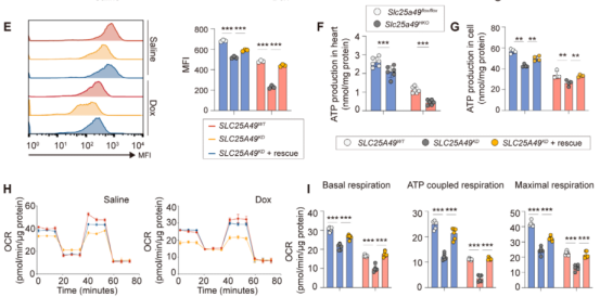
Figure 1. SLC25A49 knockdown exacerbates doxorubicin-induced mitochondrial oxidative phosphorylation dysfunction in AC16 cardiomyocytes. Image adapted from DOI: 10.1002/advs.202502163.
Journal (Impact Factor): Advanced Science (IF=14.3)
Procell product: AC16
Ⅱ. From Myocardial Ischemia to Cardioprotection and Angiogenesis
After myocardial infarction, cardiomyocyte injury, vascular dysfunction, and inflammatory responses often occur simultaneously. Determining how to intervene at different stages is an important focus of myocardial infarction research. To investigate time-dependent intervention strategies across the course of myocardial infarction, researchers used H9c2 (2-1) rat cardiomyocytes and HUVECs (human umbilical vein endothelial cells) to establish an in vitro myocardial ischemia model through oxygen-glucose deprivation (OGD). The study combined TUNEL, flow cytometry, ROS staining, Transwell assays, Matrigel tube formation assays, and qPCR for validation. The findings showed that oxygen-glucose deprivation increased reactive oxygen species (ROS) production and induced apoptosis in H9c2 (2-1) cells. Mg²⁺ reduced ROS levels and cardiomyocyte apoptosis while promoting HUVEC migration and angiogenesis.
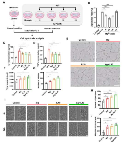
Figure 2. Regulatory effects of Mg²⁺ and IL-10 on apoptosis, angiogenesis, and inflammation in H9c2 and HUVEC cells. Image adapted from DOI: 10.1021/acsnano.6c02542.
Journal (Impact Factor): ACS Nano (IF = 16.0)
Procell products: H9c2(2-1), Human Umbilical Vein Endothelial Cells (HUVECs)
Ⅲ. From Hypertensive Cardiac Injury to Mitochondrial Protection Mechanisms
Hypertension not only increases cardiac workload but also continuously affects energy metabolism and mitochondrial function in cardiomyocytes. Could mitochondrial proteins serve as potential therapeutic targets for hypertensive heart disease? Researchers used mouse cardiomyocytes to model hypertension-related cardiac injury and established an in vitro model with L-NAME and Ang II. Through gene knockdown, overexpression, and mitochondrial function assays, they investigated the regulatory relationship among Zhx1, Mrps15, and Mrps21. The study found that the expression of Zhx1, Mrps15, and Mrps21 was altered under injury conditions. Further investigation showed that knockdown of Mrps15 or Mrps21 impaired mitochondrial function and increased ROS production, whereas Zhx1 maintained mitochondrial translation and alleviated cardiomyocyte injury by promoting Mrps15/Mrps21 expression. When Mrps15/Mrps21 were depleted, the protective effect of Zhx1 was reduced. These findings suggest that Zhx1 may protect cardiomyocytes by regulating mitochondrial ribosomal proteins and maintaining mitochondrial translation, providing new mechanistic insights for mitochondria-targeted research in hypertensive heart disease.
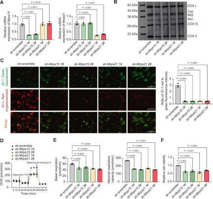
Figure 3. Mrps15/Mrps21 knockdown inhibits mitochondrial translation and induces mitochondrial dysfunction in cardiomyocytes. Image adapted from DOI: 10.1016/j.bcp.2026.118081.
Journal (Impact Factor): Biochemical Pharmacology (IF = 6.5)
Procell products: Mouse Cardiomyocytes, Mouse Cardiomyocyte Complete Medium
Ⅳ. Selected Literature Across Cardiovascular Research Areas
Beyond these areas, Procell cells and related culture products also support additional cardiovascular disease research applications. Selected literature examples are summarized below for quick reference by research area.
| Research Area | Representative Literature (Journal) | Procell Products Cited |
| Heart failure | Divergent splicing factor SRSF1 signaling promotes inflammation post-CME: the SRSF1/ENPP3 axis acts via inhibition of BRD4 O-GlcNAcylation to enhance NF-κB activation and accelerate heart failure (Theranostics, IF = 13.3) | AC16 |
| Chemotherapy-induced cardiotoxicity (multiple models) | CDC20 protects the heart from doxorubicin-induced cardiotoxicity by modulating CCDC69 degradation (Cellular & Molecular Biology Letters, IF = 9.2) | AC16, H9c2(2-1), B16-F10, Panc02, 293T [HEK-293T] |
| Viral myocarditis | Combining receptor engineering and intracellular gelation for cell-based antiviral therapy against coxsackievirus B3 (Journal of Nanobiotechnology, IF = 15.0) | HL-1, Hela |
| Myocardial infarction (targeted delivery) | Fusogenic Hybrid Gelled Nanovesicles Combining Mesenchymal Stromal Cell Cargo with Cholesterol-Modified Macrophage Membranes for Targeted Myocardial Infarction Therapy (ACS Nano, IF = 16.0) | H9c2(2-1), Human Umbilical Vein Endothelial Cells (HUVECs), RAW 264.7 |
Behind every heartbeat, countless cells work together with remarkable precision. In a culture dish, in vitro cell models recapitulate key pathological changes associated with cardiovascular disease and have become indispensable tools for cardiovascular research. From chemotherapy-induced cardiotoxicity and myocardial ischemia-reperfusion injury to hypertension-associated cardiac remodeling, appropriate and reliable cell models provide a foundation for investigating mechanisms and validating potential targets.
Prev: Choosing Serum: Look Beyond Origin and Grade to Suitability for Your Research