Exosome Purification and Characterization: Key Considerations for High-Quality Samples
Sep 21,2026
Exosome Purification and Characterization: Key Considerations for High-Quality Samples
In the previous article, we systematically reviewed the key upstream steps in exosome production, from cell source and culture system to culture mode and conditions for collecting cell culture supernatant. Once the culture supernatant is obtained, efficiently isolating the target vesicles from this complex matrix and determining whether the sample meets research requirements become critical downstream considerations.
In addition to exosomes, cell culture supernatant contains soluble contaminating proteins, lipoprotein aggregates, cellular debris, apoptotic bodies, and larger extracellular vesicles. An inappropriate purification method may result in insufficient sample purity, vesicle structural damage, and loss of biological activity, ultimately compromising functional experiments or increasing background signals in omics analyses. With the release of the MISEV2023 guidelines by the ISEV, requirements for standardized sample purification and multidimensional characterization in exosome research have become more stringent. Relying on a single purification method or a single characterization metric is often insufficient to fully support research conclusions[1]. Therefore, selecting an appropriate purification strategy and establishing a multidimensional characterization and quality control (QC) system are essential for obtaining reliable exosome samples.
Ⅰ. How Should You Choose a Purification Method? It Depends on the Separation Principle
The core principle of exosome purification is to exploit the unique physical and biochemical properties of vesicles, including their typical size range of 30–150 nm, density of 1.13–1.18 g/mL, and specific membrane markers such as CD9, CD63, and CD81, to effectively separate the target components from contaminants[2]. Currently, the major purification technologies used in basic laboratory research and clinical translation include ultracentrifugation (UC), tangential flow filtration (TFF), size-exclusion chromatography (SEC), polymer-based precipitation, and immunoaffinity chromatography. These technologies differ substantially in separation mechanism, sample recovery, purity, and ability to preserve vesicle activity and should therefore be selected according to the specific research application[3].
1. Ultracentrifugation: A Classic Method with Co-precipitation Concerns
Ultracentrifugation exploits differences in particle sedimentation behavior, using stepwise increases in centrifugal force to remove cells and debris and enrich vesicles at approximately 100,000 × g. The method requires no additional chemical reagents and is suitable for routine laboratory samples and the preliminary enrichment of relatively large volumes of supernatant. However, extremely high centrifugal forces may compress vesicles, causing membrane damage and particle aggregation. Lipoprotein complexes and soluble proteins may also co-precipitate, limiting sample purity. In addition, reproducibility between batches can be relatively poor, making the method less suitable for clinical-scale manufacturing[3].
2. TFF+SEC: Balancing Purity and Scalability
TFF concentrates samples through tangential flow circulation. Compared with conventional dead-end filtration, it can reduce membrane fouling and vesicle adsorption losses while offering good scalability for process development. Visan et al. reported that TFF+SEC outperformed UC+SEC in yield and purification efficiency, making it more suitable for processing large-volume samples[4].
Lipoproteins with particle sizes similar to those of vesicles and small inflammatory proteins that are not completely removed by TFF can be further purified by SEC. SEC relies on the molecular sieving effect of a porous gel matrix: larger exosomes cannot enter the pores and therefore elute first, whereas small contaminating proteins and free nucleic acids enter the pores and elute later. This step efficiently removes soluble contaminants and substantially improves the particle-to-total-protein ratio, making it one of the key polishing steps for improving exosome purity.
3. Polymer-Based Precipitation: Simple to Perform, but with Limited Purity
Polymer-based precipitation, particularly PEG precipitation, is simple to perform, requires minimal equipment, and generally provides relatively high recovery of various particles, making it suitable for rapid enrichment of low-concentration, small-volume samples. However, this technique relies on nonspecific co-precipitation. Along with exosomes, it can bring down substantial amounts of contaminating proteins, polysaccharides, and lipoproteins, resulting in relatively low overall sample purity. Residual PEG may also interfere with downstream experiments, including cell incubation, protein electrophoresis, and in vivo administration, potentially causing false-positive results and experimental bias[3]. Therefore, this method is better suited to preliminary exploratory experiments and should be used cautiously in formal mechanistic studies and in vivo functional studies.
4. Immunoaffinity Chromatography: High Specificity, but Limited Applications
Immunoaffinity chromatography uses immobilized antibodies against specific markers such as CD9, CD63, and CD81 to selectively capture exosome subpopulations. In theory, it provides the highest purity and enables selective isolation of specific vesicle subpopulations. However, the method is costly, elution conditions may compromise vesicle membrane integrity, and sample loss can be relatively high. It is currently used primarily for mechanistic studies of specific subpopulations and is not well suited to routine, large-scale sample preparation[2,3].
Ⅱ. How Can You Determine Whether the Purified Sample Meets Requirements?
After purification, a standardized multidimensional characterization system is essential for assessing whether an exosome sample meets quality requirements. According to the MISEV2023 guidelines, exosome studies should, where feasible, include characterization across the following dimensions: particle concentration and size distribution; morphological assessment; and protein marker profiling and contamination assessment[1]. A single characterization method is generally insufficient to support reliable research conclusions.
1. Particle-Level Characterization: Look at Size, but Pay Even More Attention to Purity
Nanoparticle tracking analysis (NTA) can be used to measure particle concentration and size distribution and is a commonly used particle-level characterization method. For exosomes derived from human Wharton’s jelly mesenchymal stem cells (hWJ-MSCs), the particle distribution within the 30–150 nm range should be evaluated together with the particle-to-total-protein ratio to assess sample purity[5].
Importantly, particle size alone cannot demonstrate that the particles are exosomes. Dynamic light scattering (DLS) has limited resolving power for complex and heterogeneous nanovesicle populations and is better suited as a complementary analytical method rather than a standalone identification criterion[1].
2. Morphological Assessment: TEM and Cryo-TEM Each Have Their Advantages
Transmission electron microscopy (TEM) can be used to observe the typical morphology and membrane structure of extracellular vesicles (such as exosomes). However, conventional negative staining, fixation, and drying procedures may alter vesicle morphology. Therefore, the commonly observed “cup-shaped” or “saucer-like” appearance does not necessarily represent the native state of exosomes and may be associated with sample preparation procedures[2].
For higher-standard basic and translational research, cryogenic electron microscopy (Cryo-TEM) can minimize the artificial deformation caused by fixation, staining, and drying, making it more suitable for observing exosomes in a state closer to their native morphology.
3. Immunodetection of Protein Markers: Both Positive and Negative Markers Are Essential
Protein marker analysis is an important step in determining vesicle origin and assessing contamination. Both positive and negative markers should be included[1].
Positive markers include the classic exosomal transmembrane proteins CD9, CD63, and CD81; ESCRT pathway-associated proteins TSG101 and Alix; and membrane raft proteins such as Flotillin[2]. Negative markers may include organelle-associated proteins such as the endoplasmic reticulum marker Calnexin. Lipoprotein markers such as APOA and APOB may also be used to assess lipoprotein contamination. Clear expression of negative markers suggests possible contamination by organelles, cell lysis products, or lipoproteins, and the effectiveness of the purification process should be further evaluated.
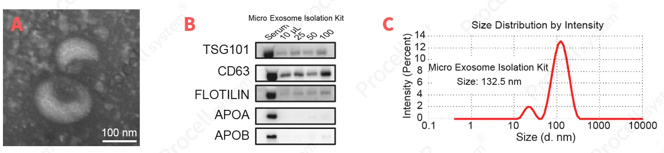
Figure 1. Characterization results of exosomes isolated using the Procell Micro Exosome Isolation Kit (Cat. No.: P-CA-501)
Ⅲ. From Basic Research to Translational Studies: QC Requirements Are Evolving
If samples are intended for animal studies or preclinical research, additional QC parameters, such as endotoxin levels, sterility, and freeze–thaw stability, should be evaluated according to the study requirements, in addition to routine analyses of particle size, concentration, morphology, and markers[1].
For samples intended for in vivo administration, endotoxin levels must be strictly controlled to avoid nonspecific inflammatory responses. Repeated freeze–thaw cycles should also be minimized to reduce the risk of vesicle membrane damage, aggregation, and loss of activity. Krishnan et al. used particle number rather than protein concentration as the basis for the dosing strategy in an in vivo safety study and systematically demonstrated that TFF-purified hWJ-MSC-derived exosomes showed no significant toxicity in vivo and were primarily distributed in the liver and spleen, providing important safety data to support clinical translation[6].
There is no single optimal exosome purification method that is suitable for every research setting. For basic research, methods such as differential ultracentrifugation can be selected according to sample volume and experimental objectives. For studies with higher requirements for purity and scalability, a TFF+SEC workflow may be considered. For clinical translation, scalable and standardized processes should serve as the foundation for establishing a systematic QC framework.
Every step in exosome research—from upstream cell culture to downstream purification and multidimensional characterization—can affect the final results. By following relevant ISEV guidelines and establishing standardized, reproducible, and traceable production and QC workflows, researchers can build a more reliable sample foundation for subsequent functional studies and clinical translation.
References
Prev: Control the Upstream Process of Exosome Production to Reduce Sample Variability