Practical Strategies for NK Cell Culture and Cryopreservation
Jun 04,2026
NK cells are key effectors of the innate immune system and play critical roles in antitumor immunity, antiviral defense, and immune regulation. Upon activation, NK cells exhibit enhanced proliferation and cytotoxicity, making them highly valuable for in vitro research and immunotherapy applications.
In this issue of Cell Culture Academy, we present practical strategies for NK cell isolation, activation, expansion, cryopreservation, and recovery to streamline and optimize experimental workflows.
I. Overview of NK Cells
NK cells are innate lymphocytes that play critical roles in immune defense. Unlike adaptive immune cells, they can directly recognize and eliminate pathogen-infected and malignant cells without prior antigen-specific stimulation.
Classical Cytotoxic Mechanisms
NK cells eliminate target cells primarily through the following cytotoxic mechanisms: (Figure 1)
1.Perforin-Granzyme Pathway
NK cells release cytotoxic granules containing perforin and granzymes. Perforin forms pores in the target cell membrane, enabling granzymes to enter the cell and trigger apoptosis or apoptosis.
2.Fas-FasL Death Receptor Pathway
Fas ligand (FasL) on NK cells binds to Fas receptors on target cells, activating apoptotic signaling pathways that induce target cell apoptosis.

Figure 1. Schematic of NK Cell Cytotoxic Mechanisms.
(Adapted from Concise Clinical Immunology for Healthcare Professionals)
Phenotypic Characteristics and Subsets
NK cells are classically defined by the CD3⁻ CD56⁺ phenotype, with CD3 used to distinguish them from CD3⁺ CD56⁺ NKT cells.
Based on CD56 expression, NK cells are divided into two functionally distinct subsets:
CD56bright NK cells: Express high levels of CD56 and low or no CD16.
These cells primarily mediate immunoregulatory functions through secretion of cytokines such as IFN-γ and TNF-α. They exhibit strong proliferative capacity but relatively weak cytotoxicity, contributing mainly to early antiviral responses and immune homeostasis.
CD56dim NK cells: Express low levels of CD56 and high levels of CD16.
These cells display potent cytotoxic activity with comparatively limited cytokine secretion. Their effector functions are primarily mediated through natural cytotoxicity and CD16-dependent antibody-dependent cellular cytotoxicity (ADCC).
II. Isolation and Culture of NK Cells
The isolation and culture procedures for umbilical cord blood-derived NK cells are largely similar to those for peripheral blood-derived NK cells. However, because cord blood contains a lower baseline frequency of NK cells, a longer expansion period is typically required to achieve sufficient cell yields. The following protocol uses peripheral blood-derived NK cells as an example.
1. Isolation of Peripheral Blood Mononuclear Cells (PBMCs)
Centrifuge fresh whole blood at 2,300 rpm for 10 min. Collect the plasma layer and dilute it 1:1 with PBS.
Carefully overlay the diluted blood onto Ficoll separation medium and centrifuge at 2,000 rpm for 20 min with low acceleration (setting 1) and no brake.
From top to bottom, the layers appear as follows: the plasma layer, cell layer, separation medium layer, and red blood cell/granulocyte layer. The cell layer contains the PBMC fraction (Figure 2).
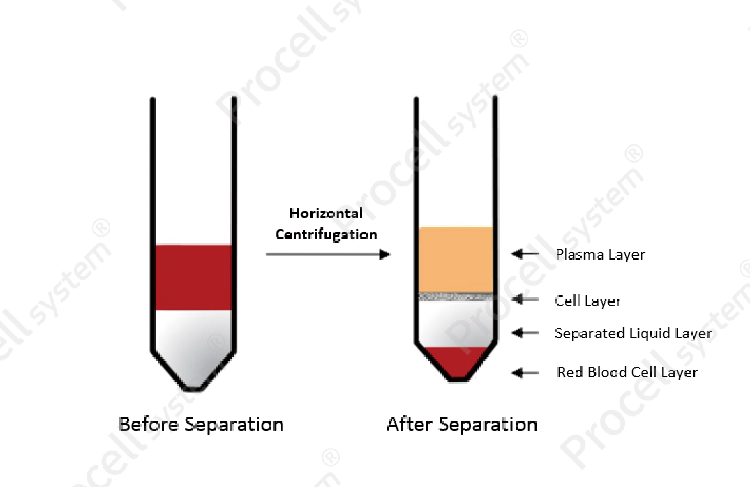
Figure 2. Schematic illustration of PBMC isolation by Ficoll density gradient centrifugation.
Carefully transfer the PBMC layer to a new centrifuge tube. Wash the cells 2-3 times with PBS by centrifugation at 1,200 rpm for 8 min each using low acceleration (setting 1) and no brake.
Resuspend the cells in PBS and determine cell density by cell counting.
2. NK Cell Activation and Culture
Coat the culture vessel with a soluble anti-CD16 antibody and incubate overnight at 4℃.
Wash the coated vessel 2-3 times, then add serum-free medium supplemented with cytokines such as IL-2 and IL-15.
Seed PBMCs at a density of 2-2.5 × 10⁶ cells/mL in serum-free medium and culture at 37℃ in a humidified 5% CO₂ incubator.
3. NK Cell Expansion Culture
During the first week of culture, 5% heat-inactivated autologous plasma may be added to enhance cell viability and proliferation.
Replenish the culture every 2 days with fresh serum-free medium and cytokines required for NK cell growth. Monitor cell density regularly and maintain it at approximately 1 × 10⁶ cells/mL to support optimal proliferation.
4. NK Cell Characterization
NK cells are primarily characterized by flow cytometric analysis of surface markers (CD3⁻ CD56⁺), combined with functional assays to evaluate cytotoxic activity.
Cytotoxicity is commonly assessed using effector-to-target (E:T) ratio assays, in which NK cells are co-cultured with target cells at different ratios, and target cell lysis is measured to quantify NK cell killing activity.
III. Cryopreservation and Recovery of NK Cells
1. Cryopreservation
Use cryopreservation medium containing 10% DMSO to protect cell membranes during low-temperature storage. Resuspend harvested NK cells in freezing medium at a density of 1 × 10⁶-1 × 10⁷ cells/mL.
Transfer the cell suspension into cryovials and perform controlled-rate freezing. Store the cryovials at -80℃ overnight before transferring them to liquid nitrogen for long-term preservation.
For serum-free applications, serum-free cryopreservation medium may be used.
2. Recovery
Prewarm the water bath to 37℃ and equilibrate the culture medium before thawing.
Remove the cryovial from storage and immediately thaw it in a 37℃ water bath with gentle agitation.
Transfer the thawed cells into a 15 mL centrifuge tube containing 5 mL prewarmed culture medium. Centrifuge at 300 × g for 5 min and discard the supernatant.
Resuspend the cells in complete medium, transfer them to a prepared culture vessel, and incubate at 37℃ in a humidified 5% CO₂ incubator.
IV. Experimental Data Presentation
Human NK cells expanded using antibody-cytokine stimulation exhibited typical cluster-like growth morphology (Figure 3). Flow cytometry showed that the CD3⁻ CD56⁺ population exceeded 95% (Figure 4). NK cells also demonstrated potent cytotoxicity against K562 cells across multiple effector-to-target (E:T) ratios (Figure 5).
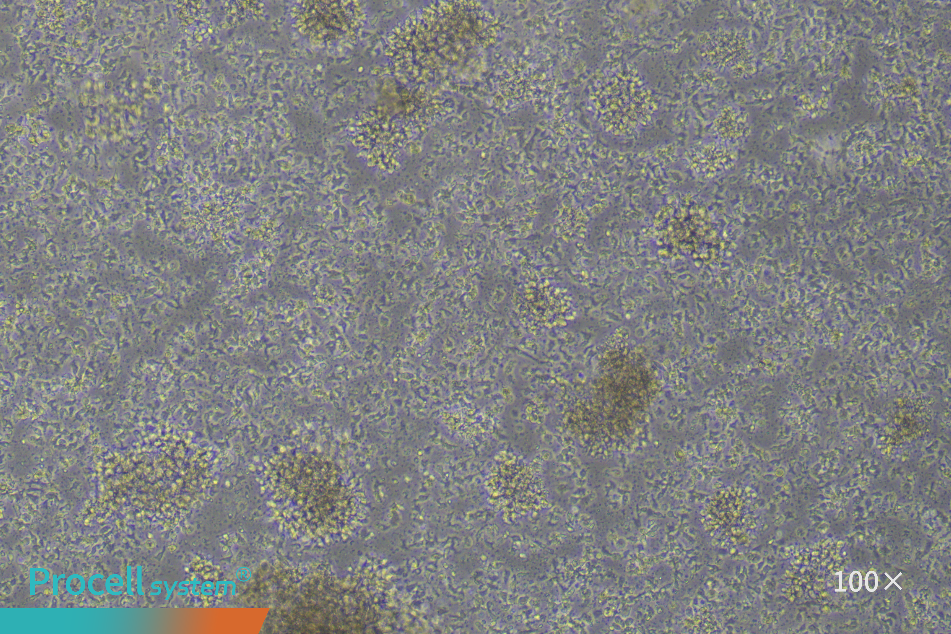
Figure 3. Bright-field microscopic image of NK cells
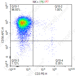
Figure 4. Flow cytometric analysis of CD3 and CD56 expression in expanded NK cells

Figure 5. Quantification of NK cell cytotoxicity at different E:T ratios
Prev: Optimizing in Vitro T Cell Culture: A Step-by-Step Experimental Protocol

