Troubleshooting Hepatocyte and Non-parenchymal Cell Isolation: Essential Tips for Successful Cell Preparation
Aug 05,2026
The liver is the largest internal organ involved in metabolism and digestion and is located in the upper right region of the abdomen. It performs essential physiological functions, including metabolism, detoxification, coagulation, and immune regulation. Due to its diverse cell populations, complex tissue architecture, extensive vascularization, and cellular heterogeneity, the liver presents significant challenges for the isolation of high-quality primary cells.
In this issue of Cell Culture Academy, we summarize key techniques for the isolation and culture of hepatic parenchymal and non-parenchymal cells, providing researchers with a comprehensive overview of primary liver cell isolation strategies and approaches for experimental optimization.
I. Overview of Liver Cell Types
Liver cells are broadly categorized into two major groups: hepatic parenchymal cells and hepatic non-parenchymal cells (NPCs) (Figure 1).
● Hepatic Parenchymal Cells (Hepatocytes)
Hepatocytes constitute approximately 70–80% of the liver cell population and represent the major cellular component of the liver. These large, typically polygonal cells often exhibit a characteristic binucleated morphology and perform essential liver functions, including metabolism, detoxification, and bile secretion. Due to their central role in hepatic function, hepatocytes are widely used as a primary cellular model for investigating liver physiology, toxicology, and disease mechanisms.
● Hepatic Non-Parenchymal Cells (non-parenchymal cells)
Hepatic non-parenchymal cells account for approximately 20–30% of total liver cells and play essential roles in immune regulation, intercellular communication, and maintenance of liver homeostasis. The major hepatic non-parenchymal cell populations include:
Hepatic Stellate Cells (HSCs)
HSCs are key regulators of hepatic extracellular matrix remodeling. In the healthy liver, they serve as the primary storage site for vitamin A. Following liver injury, activated HSCs contribute to tissue repair and fibrogenesis, making them widely used models for studying liver fibrosis, regeneration, and tissue remodeling.
● Kupffer Cells
Kupffer cells are specialized liver-resident macrophages with important phagocytic and clearance functions. As the largest resident immune cell population in the liver, they play critical roles in hepatic immune surveillance, inflammation, and host defense. Kupffer cells are widely utilized in studies of hepatic inflammation, infection, and immune regulation.
● Liver Sinusoidal Endothelial Cells (LSECs)
LSECs constitute the specialized endothelial lining of hepatic sinusoids and form a critical vascular interface within the liver. Their fenestrated structure facilitates efficient exchange of plasma components and macromolecules between blood and the hepatic parenchyma. LSECs are essential regulators of the hepatic immune microenvironment and serve as important models for studying vascular biology, immune regulation, and transmembrane transport mechanisms.
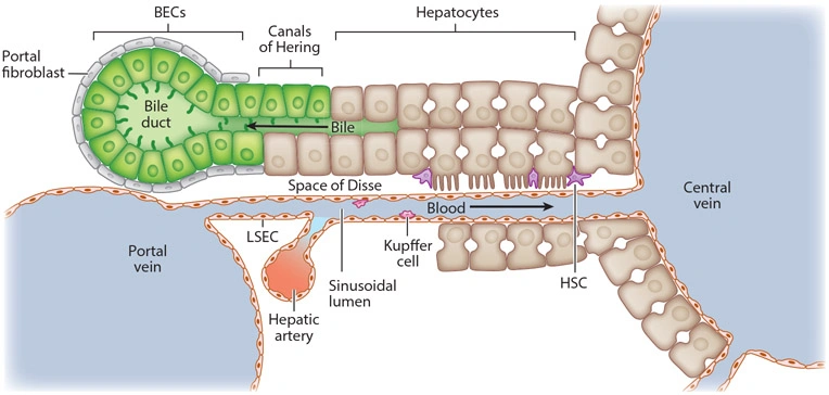
Figure 1. Schematic illustration of the locations of hepatic parenchymal and non-parenchymal cells(Image adapted from the literature: Multiple Facets of Cellular Homeostasis and Regeneration of the Mammalian Liver)
II. Challenges in Liver Cell Isolation
The liver is a highly vascularized parenchymal organ with a complex cellular architecture. Hepatic parenchymal cells and non-parenchymal cells are densely interspersed throughout the tissue, along with abundant hepatic sinusoids and extracellular matrix components, which collectively increase the complexity of liver tissue dissociation. Compared with tissues such as the spleen and thymus, which contain relatively loose connective structures, liver cell isolation requires precise control and optimization of enzymatic digestion conditions.
Hepatocytes are tightly arranged along hepatic plates. Mechanical dissociation alone can result in significant cellular damage and increased contamination by blood cells and tissue debris, thereby compromising cell yield, purity, and viability. Therefore, achieving efficient tissue dissociation while maintaining cellular integrity represents a major challenge in hepatocyte isolation.
Hepatic non-parenchymal cells (NPCs) account for only 20–30% of total liver cells, with individual populations, including hepatic stellate cells (HSCs), Kupffer cells, and liver sinusoidal endothelial cells (LSECs), representing an even smaller fraction. Due to their comparable cell density and in vitro adhesion properties, different NPC populations are prone to cross-contamination during isolation. Therefore, the implementation of appropriate enrichment and purification strategies is essential for obtaining highly purified hepatic non-parenchymal cell populations.
III. Isolation Methods for Different Liver Cell Types
1. Hepatocytes
Hepatocyte isolation is commonly performed using the two-step collagenase perfusion method (Figure 2). Briefly, prewarmed buffer is first perfused through the portal vein in anesthetized animals to remove blood and circulating cells, followed by collagenase perfusion to enzymatically digest the liver tissue under controlled conditions. After sufficient digestion, the liver is gently dissociated to generate a single-cell suspension, which is filtered through a cell strainer and subjected to low-speed centrifugation to remove tissue debris.
The resulting cell suspension is further purified by density gradient centrifugation to enrich highly viable hepatocytes. Isolated hepatocytes are initially cultured in an attachment-promoting medium to enhance cell attachment and improve early cell survival. Once stable attachment is achieved, the medium is replaced with maintenance medium for short-term recovery culture to preserve hepatocyte-specific phenotypes and functional characteristics while minimizing dedifferentiation during in vitro culture. This process enables the preparation of highly enriched and viable hepatocyte populations.
Successful hepatocyte isolation largely depends on proper portal vein cannulation and efficient perfusion. Before conducting the actual experiment, it is recommended to practice the perfusion procedure using PBS in experimental animals.
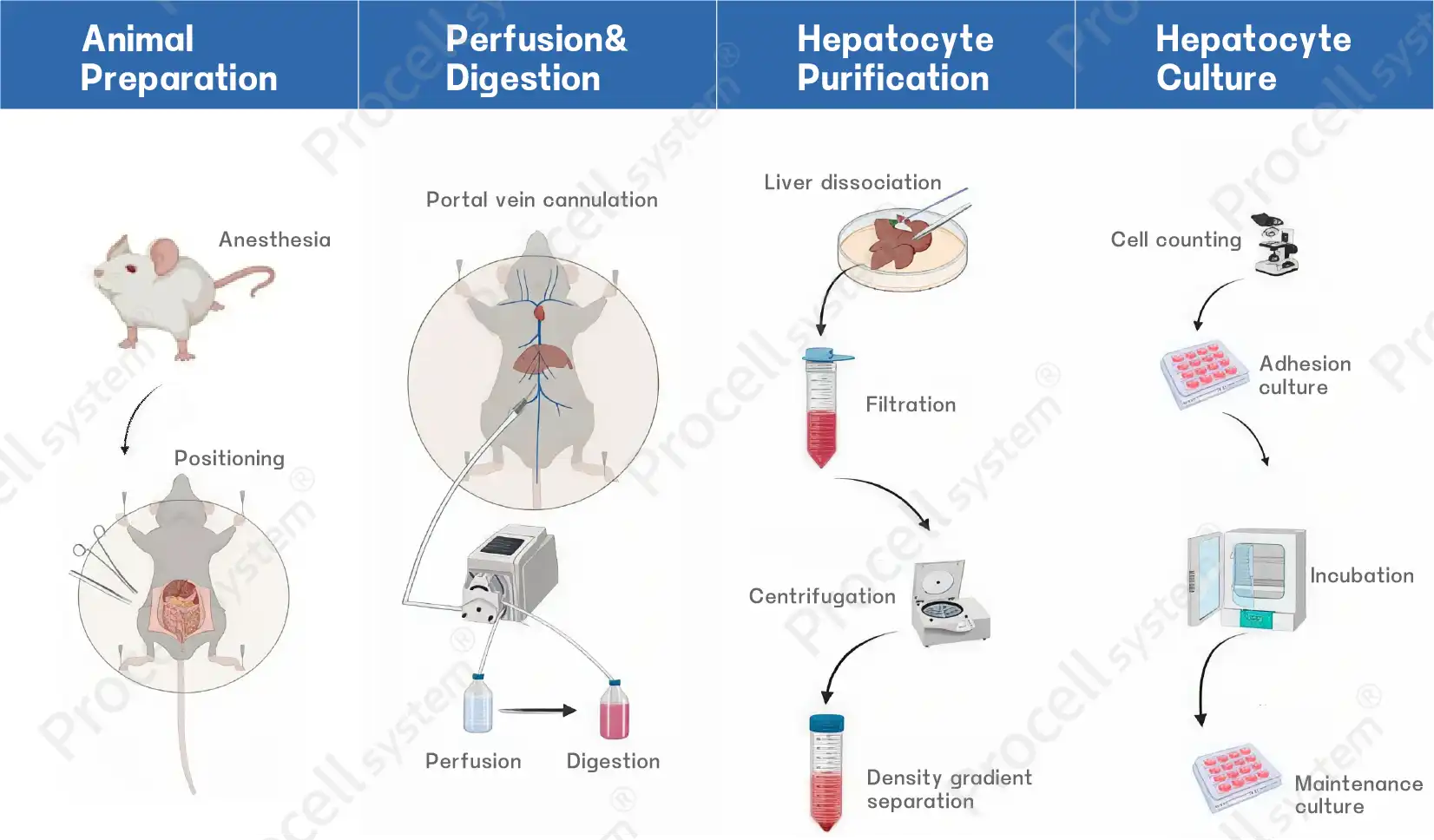
Figure 2. Schematic diagram of hepatocyte isolation
2. Kupffer Cells and Liver Sinusoidal Endothelial Cells
The standard isolation procedure for Kupffer cells and liver sinusoidal endothelial cells involves mechanical dissociation of liver tissue, overnight enzymatic digestion, and generation of a single-cell suspension. The target cell populations are initially enriched by density gradient centrifugation. However, a single density gradient separation step is insufficient to achieve complete separation of these two cell types. Therefore, a secondary purification step based on differences in cell adhesion properties is commonly performed using the differential adhesion method.
A typical differential adhesion-based isolation procedure (using rat liver tissue as an example) is shown in Figure 3. This method utilizes the different adhesion rates of Kupffer cells and LSECs. The target cell population enriched by density gradient centrifugation is seeded into culture dish A and incubated for 20-25 min. During this period, the faster-adhering Kupffer cells attach to the dish surface, whereas LSECs remain in the suspension fraction.
● Isolation of LSECs:
The suspension fraction from culture dish A is gently collected and transferred to a new culture dish (dish B) for further culture. The resulting cell population in dish B is enriched in LSECs.
● Isolation of Kupffer cells:
The adherent cells remaining in culture dish A are Kupffer-enriched fraction. After removal of the supernatant, fresh complete medium is added, and the medium is replaced 1–2 additional times to further remove non-adherent contaminating cells.
It should be noted that the optimal differential adhesion time may vary depending on the species and experimental conditions. For optimal results, the recommended parameters in the product-specific protocol should be followed.
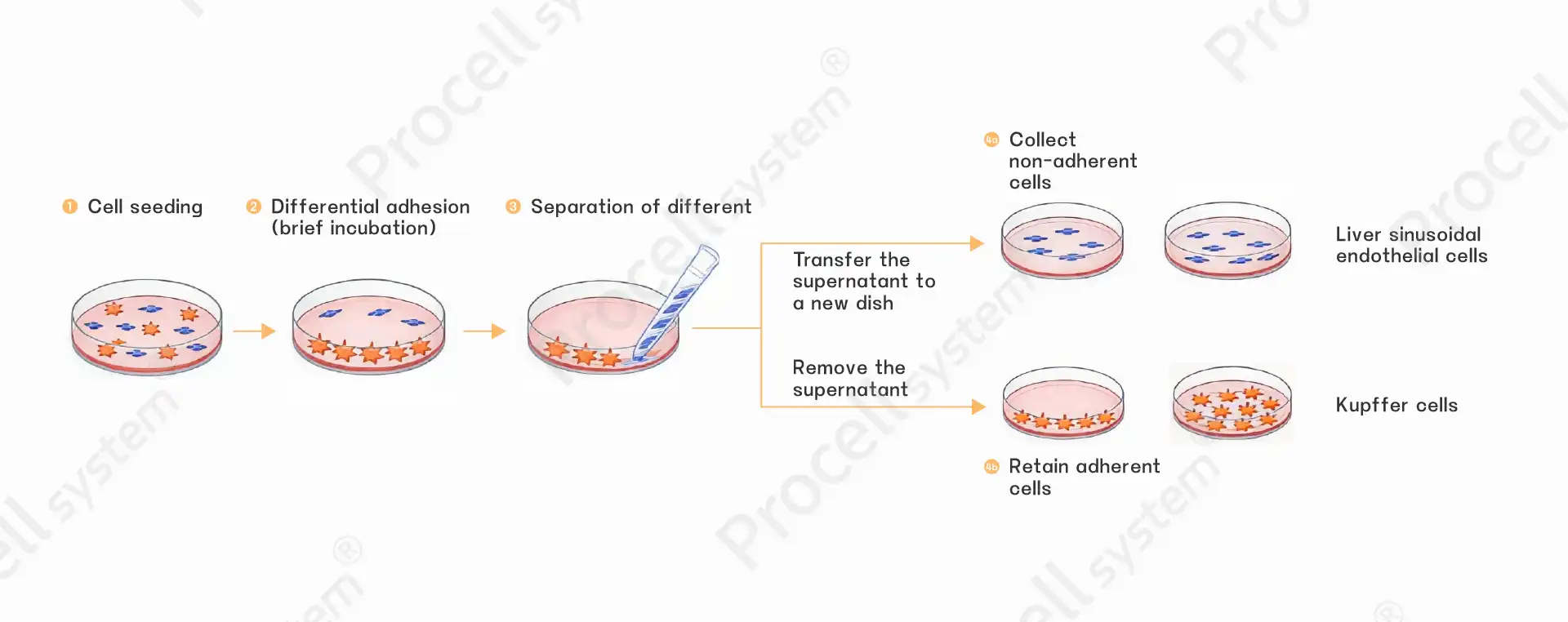
Figure 3. Schematic diagram of the differential adhesion process
3. Hepatic Stellate Cells
The isolation procedure for hepatic stellate cells (HSCs) is relatively simple. Briefly, liver tissue is mechanically dissociated and enzymatically digested for 30–40 min to generate a single-cell suspension, followed by density gradient centrifugation to enrich the target cell population. The isolated cells are then cultured under HSC-supportive culture conditions to promote cell attachment and maintenance, resulting in highly pure and viable HSCs.
Compared with Kupffer cells and liver sinusoidal endothelial cells, HSC isolation is highly influenced by the age of the donor animals, and neonatal rodents are commonly used. In addition, this procedure does not require overnight digestion, which helps minimize cell damage and improve cell viability.
IV. Key Considerations for Liver Cell Culture
Table 1 summarizes the morphological characteristics, culture properties, passaging capacity, and commonly used digestion methods of hepatocytes and non-parenchymal liver cells (NPCs), providing a reference for selecting appropriate culture conditions.
Table 1. Cell Culture Parameters for Liver Cells
|
Cell Type |
Morphological Characteristics |
Culture Characteristics |
Passaging Capacity |
Digestion Enzymes |
|
Hepatocytes |
Binucleated, large polygonal cells |
Rapid dedifferentiation after attachment; sensitive to culture conditions |
Non-passagable |
Not applicable |
|
Kupffer Cells |
Spindle-shaped, macrophage-like morphology |
Rapid attachment; strong phagocytic activity |
Non-passagable |
Pronase (12 mM) / 0.25% Trypsin |
|
Hepatic Stellate Cells (HSCs) |
Fibroblast-like morphology |
Gradual activation after attachment; relatively strong proliferative capacity |
Passagable; optimal phenotype maintained within 3 passages |
0.25% Trypsin |
|
Liver Sinusoidal Endothelial Cells |
Cobblestone-like morphology |
Sensitive to culture conditions; prone to phenotypic changes |
Passagable for 1–2 passages |
0.25% Trypsin |
For more information on primary cell isolation solutions, please click Primary Cell Isolation Kits to explore related products.
Prev: Direct vs Indirect vs 3D Co-culture: Selecting the Right Model for Your Research
Next: Essential Guide to THP-1 Cell Culture, Differentiation, and Macrophage Polarization