Essential Guide to THP-1 Cell Culture, Differentiation, and Macrophage Polarization
Aug 05,2026
Macrophages are key effector cells of the innate immune system and play important roles in inflammatory regulation, immune responses, and tissue repair. Due to their high functional plasticity, macrophages are widely used as model systems for studying immune regulation mechanisms. However, macrophage phenotypes are highly influenced by microenvironmental conditions, making the establishment of stable and reproducible in vitro models essential.
The human monocytic leukemia cell line THP-1 exhibits typical monocyte characteristics and can be induced to differentiate into macrophage-like cells under specific conditions. Therefore, THP-1 cells are widely used in studies of monocyte-to-macrophage differentiation and macrophage function. This guide summarizes the principles and key considerations involved in THP-1 cell culture, differentiation, and polarization.
1. Introduction to THP-1 Cells
THP-1 (human monocytic leukemia cells) was established in 1980 by Tsuchiya's group at Tohoku University, Japan, from the peripheral blood of a 1-year-old child diagnosed with acute monocytic leukemia.
Compared with primary human peripheral blood mononuclear cells (PBMCs), THP-1 cells offer several advantages:
• Stable source: As an immortalized cell line, THP-1 cells can be continuously passaged, reducing experimental variability caused by donor differences during primary cell isolation.
• Defined differentiation potential: THP-1 cells retain key functional characteristics of primary monocytes, including phagocytosis, lysozyme activity, and the ability to respond to environmental signals for differentiation and polarization.
• Mature culture system: Standardized culture and induction protocols are available, making THP-1 suitable for mechanistic studies and drug screening.
2. THP-1 Cell Culture and Maintenance
Maintaining stable cell status is critical for reproducible differentiation and polarization experiments.
Culture conditions
Recommended culture medium: RPMI-1640 + 10% FBS + 0.05 mM β-mercaptoethanol + 1% penicillin/streptomycin
Incubation conditions:
• Atmosphere: 95% air, 5% CO₂
• Temperature: 37°C
Passaging: THP-1 cells can be maintained by adding fresh medium or by centrifugation and medium replacement. A recommended centrifugation condition is 1,200 rpm (approximately 250×g) for 3 min.
Medium replacement frequency: 2-3 times per week.
Key maintenance considerations
• Maintain appropriate cell density. THP-1 cells are suspension cells and are sensitive to density changes. Low density may result in slow proliferation and poor cell status, while excessive density may cause nutrient depletion. A density of 5×10⁵–1×10⁶ cells/mL is generally recommended.
• Recovery after thawing. THP-1 cells are relatively difficult to recover after cryopreservation. During the first week after thawing, gentle culture methods such as medium supplementation or partial medium replacement are recommended to minimize mechanical stress.
• Maintain a mildly acidic growth environment. Phenol-red-containing RPMI-1640 medium appears orange-yellow at pH approximately 6.8–7.2. A red or purple color indicates elevated pH, which may affect cell growth.
• Add β-mercaptoethanol. β-mercaptoethanol maintains a reducing microenvironment and is important for stabilizing THP-1 physiological characteristics.
• Serum quality affects cell performance. Poor-quality serum may cause enlarged cells, aggregation, or abnormal attachment. Validated serum lots and consistent culture conditions are recommended.
3. Principles of THP-1 Differentiation and Macrophage Polarization
In vivo, circulating monocytes migrate from blood into tissues in response to chemotactic signals and differentiate into resting M0 macrophages under local microenvironmental regulation.
M0 macrophages are highly plastic and can be further polarized into different functional states
• M1 macrophages mainly participate in pro-inflammatory responses and pathogen clearance.
• M2 macrophages are primarily involved in immune regulation, inflammation resolution, and tissue repair.
Polarization mechanisms
M1 polarization:
LPS activates TLR4 signaling and NF-κB pathways, while IFN-γ activates JAK-STAT1 signaling. These pathways promote expression of pro-inflammatory genes and drive M1 polarization.
M2 polarization:
IL-4 and IL-13 activate the JAK-STAT6 pathway, promoting STAT6 phosphorylation and expression of anti-inflammatory and tissue repair-related genes.
In vitro THP-1 differentiation model
Untreated THP-1 cells normally grow as suspended monocytes. PMA, a protein kinase C (PKC) activator, mimics differentiation signals and induces THP-1 cells to transition into adherent macrophage-like cells. Following PMA stimulation, THP-1 cells stop proliferating and mature into M0 macrophages, which can then be polarized into M1 or M2 macrophages using specific cytokines.
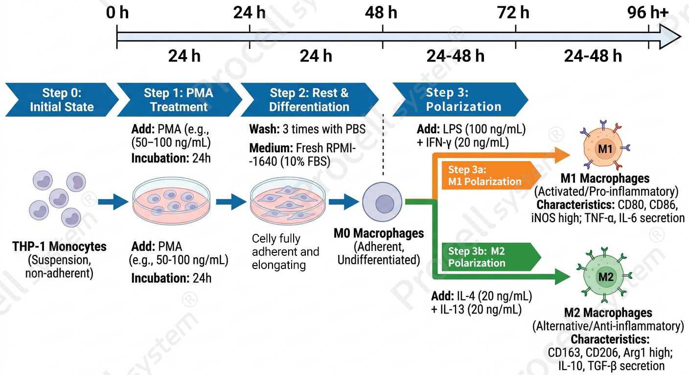
Figure 1. Workflow of THP-1 cell differentiation and macrophage polarization
4. THP-1 In Vitro Differentiation and Polarization Workflow
M0 macrophage induction
Seed healthy THP-1 cells at 5×10⁵ cells/mL and add PMA at a final concentration of 50–100 ng/mL. Incubate for 24 h.
After induction, remove PMA-containing medium and replace with PMA-free complete medium. Continue incubation for another 24 h to allow recovery and formation of stable adherent macrophage-like cells.
Morphological characteristics:
Untreated THP-1 cells: round morphology, suspension growth, smooth cell surface.
PMA-induced M0 macrophages: gradual attachment, cell spreading, and macrophage-like morphology.
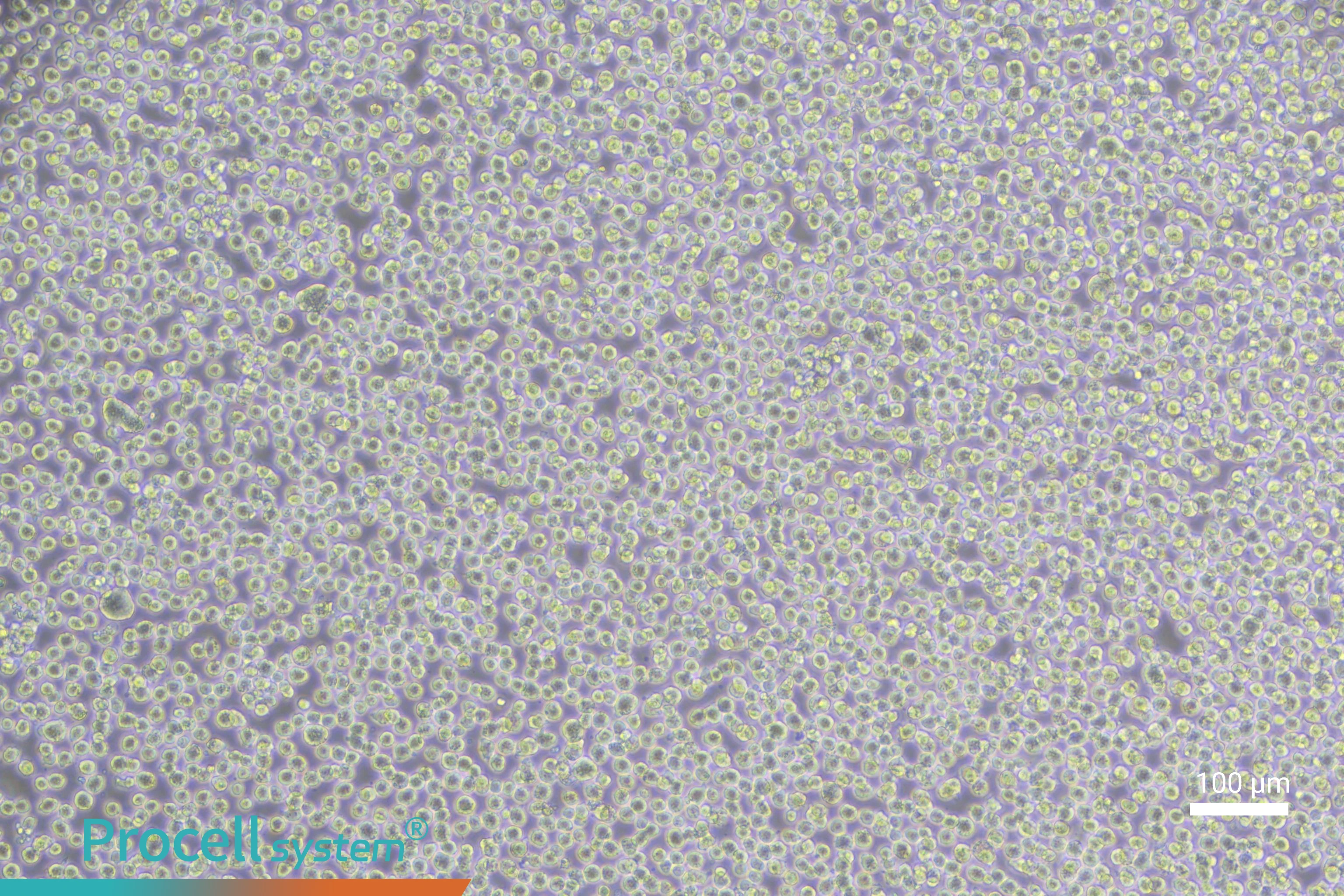
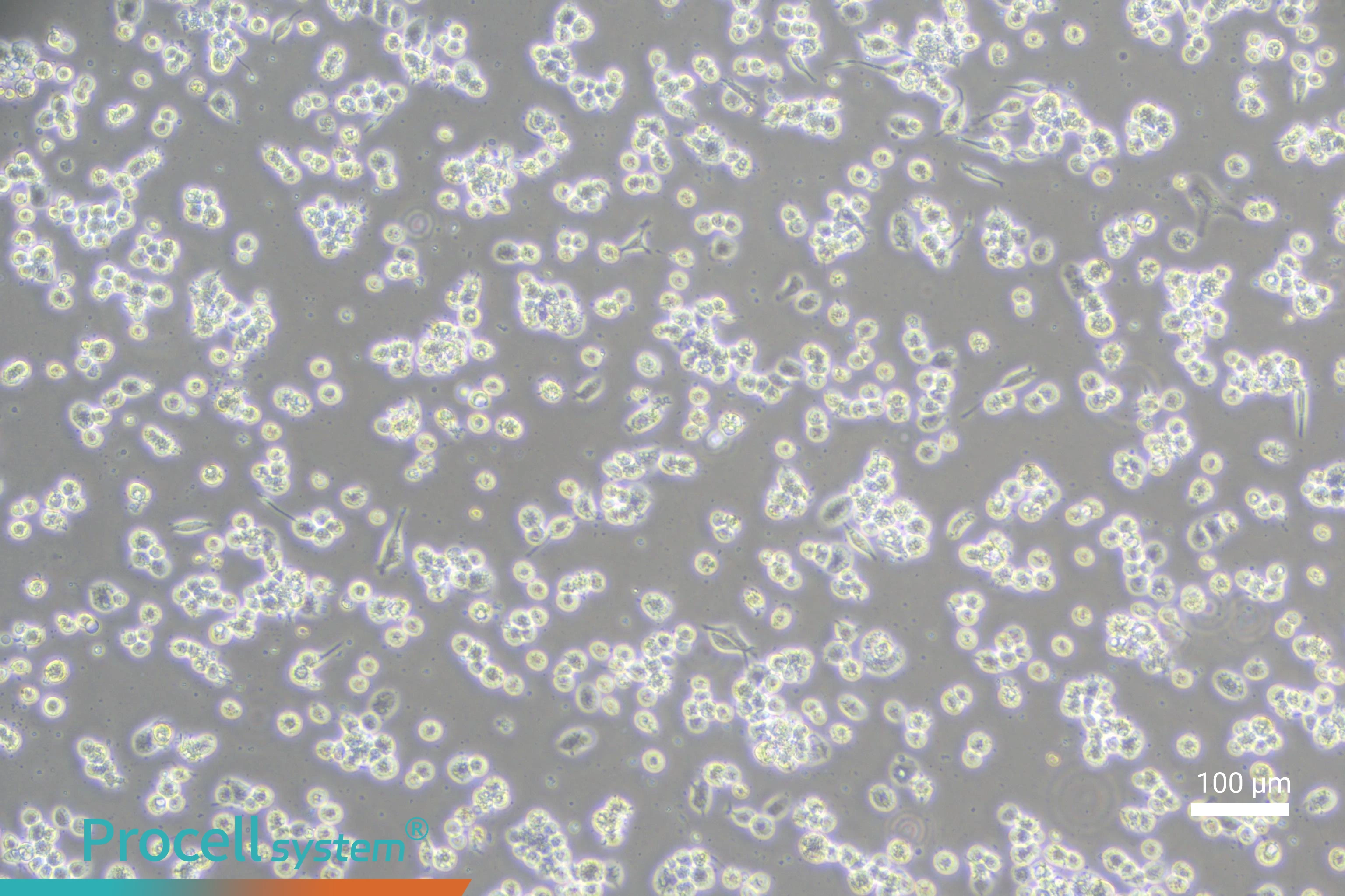
Figure 2. Morphological comparison of THP-1 cells and M0 macrophages (left: THP-1 cells; right: M0 macrophages)
M1 polarization induction:
Add LPS (100 ng/mL) and IFN-γ (20 ng/mL) to M0 macrophage cultures and incubate for 24–48 h.
Typical M1 characteristics:
Cells become larger, polygonal, and spread with extended filopodia. Increased vacuole formation may occur due to enhanced phagocytic activity.
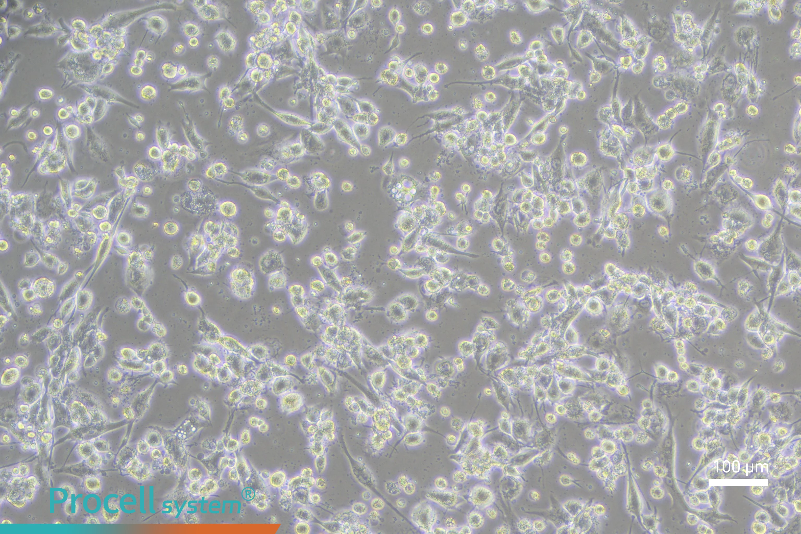
Figure 3. Morphology of M1 macrophages
M2 polarization induction
Add IL-4 (20 ng/mL) and IL-13 (20 ng/mL; IL-10 can also be used as an alternative) and incubate for 24–48 h.
Typical M2 characteristics:
Cells are generally smaller and more compact, with round or spindle-like morphology, fewer protrusions, denser cytoplasm, and more organized arrangement.
Validation of Polarization Status
Common validation methods include:
• qPCR analysis: Detect M1 markers TNF-α and IL-1β, and M2 markers CD206 and Arg1 at the mRNA level.
• Flow cytometry: Detect M1 markers CD80/CD86 and M2 markers CD206/CD163 at the protein level.
• ELISA: Measure cytokine secretion levels such as TNF-α and IL-10 in culture supernatants.
5. Common Problems and Optimization Strategies
|
Problem |
Possible Cause |
Optimization Strategy |
|
Poor cell attachment |
Insufficient PMA concentration |
Increase PMA concentration appropriately (not exceeding 100 ng/mL) |
|
Suboptimal cell growth status |
Use THP-1 cells in the logarithmic growth phase with good viability |
|
|
Uneven differentiation (mixed suspension and adherent cells with obvious morphological differences) |
Cell suspension not thoroughly mixed before seeding |
Mix the cell suspension thoroughly before induction to ensure uniform cell distribution |
|
Inappropriate seeding density |
Optimize cell seeding density and avoid excessively high or low densities |
|
|
Poor polarization efficiency (weak marker expression) |
Residual PMA affects subsequent polarization |
Completely remove PMA after differentiation and replace with fresh medium |
|
Insufficient activity of induction factors |
Use freshly prepared cytokines or cytokines with validated activity |
Next: Cardiac Primary Cell Isolation: Key Considerations for Three Major Cell Types