Cardiac Primary Cell Isolation: Key Considerations for Three Major Cell Types
Aug 26,2026
The heart is the central organ of the vertebrate circulatory system. Through rhythmic contraction and relaxation, it continuously drives systemic blood circulation and maintains normal physiological functions.
Cardiac tissue contains multiple cell populations that play essential roles in cardiac development, homeostasis, tissue repair, and disease progression. Therefore, cardiac cell isolation and culture are important foundations for cardiovascular disease research, regenerative medicine, drug screening, and tissue engineering.
However, primary cardiac cell isolation involves complex procedures and multiple influencing factors. Common challenges include low cell yield, poor viability, insufficient purity, and loss of cellular characteristics after limited passages. This article summarizes the key principles and technical considerations for isolating and culturing three major cardiac cell types.
Ⅰ. Major Cardiac Cell Types for Research
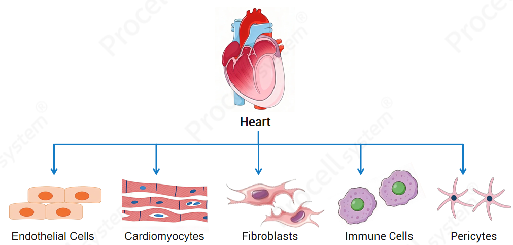
Cardiac tissue consists of multiple cell populations, including endothelial cells, cardiomyocytes, cardiac fibroblasts, immune cells, and pericytes. Among these, cardiomyocytes, cardiac fibroblasts, and cardiac microvascular endothelial cells (CMECs) are the most commonly studied.
Cardiomyocytes
Cardiomyocytes are the major functional cells of the heart. They exhibit spontaneous contraction and possess four key physiological properties: excitability, automaticity, conductivity, and contractility, which are essential for maintaining cardiac rhythm and pumping function.
Cardiac Fibroblasts
Cardiac fibroblasts are the most abundant non-myocyte cells in the heart. They mainly produce extracellular matrix (ECM) components such as collagen. After cardiac injury, fibroblasts become activated and contribute to scar formation. Persistent activation may lead to cardiac fibrosis and impaired cardiac function.
Cardiac Microvascular Endothelial Cells
Cardiac microvascular endothelial cells exhibit a typical cobblestone-like morphology and are important components of the cardiac microcirculation. They regulate vascular barrier function, vascular tone, inflammatory responses, leukocyte recruitment, and angiogenesis.
Ⅱ. Five Major Steps in Primary Cardiac Cell Isolation
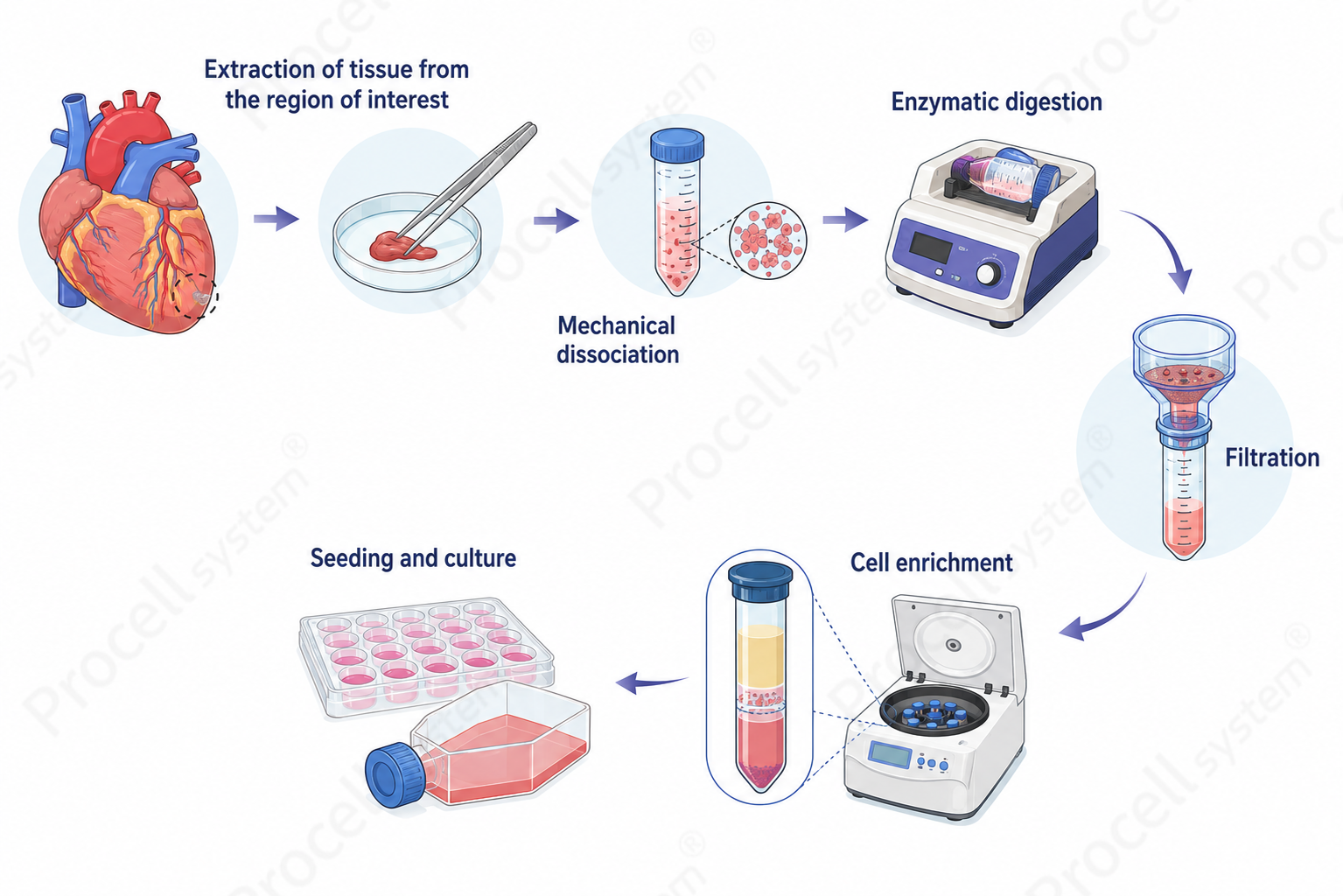
The general workflow includes five major steps: tissue collection and pretreatment, enzymatic digestion, filtration, cell purification and enrichment, and cell seeding and culture.
1. Tissue Collection and Pretreatment
Cardiac tissue is collected under sterile conditions. Blood and connective tissues are removed, and appropriate tissue regions are selected according to the target cell type.
2. Enzymatic Digestion
Collagenase, trypsin, or other digestive enzymes are used to degrade extracellular matrix components and release target cells. Enzyme type, concentration, and digestion time should be optimized to balance efficiency and viability.
3. Filtration and Debris Removal
The digested suspension is filtered through a 70–100 μm cell strainer to remove tissue debris and obtain a relatively uniform single-cell suspension.
4. Cell Purification and Enrichment
Common purification strategies include differential adhesion, density gradient centrifugation, and selective drug-based purification.
5. Cell Seeding and Culture
Purified cells are seeded into culture vessels and maintained using optimized culture systems specific to each cell type.
Ⅲ. Key Differences Among Three Cardiac Cell Types
Different cardiac cell types require different strategies for animal age selection, purification, culture conditions, and passaging.
1. Animal Age Selection
Commonly used experimental animals include KM, C57BL/6, and BALB/c mice, as well as Sprague-Dawley (SD) and Wistar rats.
Animal age is an important factor affecting cell yield, viability, and culture success. The recommended ages vary depending on the target cell type as follows:
Table 1. Recommended Animal Age Selection for Isolation of Different Cardiac Primary Cells
|
Cell Type |
Recommended Age |
Rationale |
|
Cardiomyocytes |
1–2 days old (mice or rats) |
Neonatal cardiomyocytes show stronger tolerance to enzymatic digestion and higher viability and attachment rates |
|
Cardiac fibroblasts |
1–2 days old (mice or rats) |
Neonatal cardiac fibroblasts exhibit stronger proliferative capacity and higher culture success rates |
|
Cardiac microvascular endothelial cells |
15–20 days old (mice or rats) |
More mature cardiac microvascular development facilitates microvascular segment isolation and endothelial cell recovery |
2. Significant Differences in Purification Strategies
The biological characteristics of different cardiac cell types determine the appropriate purification methods, resulting in substantial differences among cell isolation strategies.
Cardiomyocytes and Cardiac Fibroblasts
The major difference between these two cell types lies in their attachment kinetics. Under typical conditions, cardiac fibroblasts attach within approximately 30 min, whereas cardiomyocytes generally require approximately 2 h for attachment. Therefore, differential adhesion can be used for preliminary separation.
In addition, cardiac fibroblasts exhibit strong proliferative capacity, while cardiomyocytes have limited proliferative ability. Therefore, selective purification strategies, such as the use of proliferation inhibitors, can be applied during culture to further improve the purity of cardiomyocyte populations.
Cardiac Microvascular Endothelial Cells
Cardiac microvascular endothelial cells are typically isolated from microvascular fragments within cardiac tissue. During culture, endothelial cells gradually migrate out from these microvascular explants.
Because microvascular fragments have different density characteristics compared with other tissue components, density gradient centrifugation is commonly used to enrich microvascular fragments, thereby improving the efficiency of endothelial cell isolation.
3. Differences in Culture and Passaging Methods
Different cardiac cell types exhibit distinct characteristics in terms of morphology, growth patterns, and passaging conditions.
Table 2. Comparison of Culture Characteristics and Passaging Conditions of Cardiac Primary Cells
|
Cell Type |
Morphological Features |
Culture Characteristics |
Recommended Dissociation Reagent |
|
Cardiomyocytes |
Spindle-shaped or polygonal morphology; spontaneous contraction may be observed |
Gradually extend after culture and form synchronously beating cell clusters; generally not suitable for passaging |
|
|
Cardiac fibroblasts |
Typical fibroblast-like morphology |
Rapid attachment and strong proliferation ability; suitable for limited passages |
|
|
Cardiac microvascular endothelial cells (CMECs) |
Cobblestone-like morphology |
Gradually migrate from microvascular segments; low cell yield during early culture; commonly passaged for 2–3 generations |
Prev: Essential Guide to THP-1 Cell Culture, Differentiation, and Macrophage Polarization
Next: Serum-Containing vs. Serum-Free – Choosing the Right Cell Culture System