Adherent Cell Dissociation: Selection and Optimization of Reagents and Digestion Endpoints
Jun 12,2026
Dissociating and passaging adherent cells is a routine yet critical step in cell culture. Improper handling can reduce viability and proliferation, trigger apoptosis, and compromise experimental outcomes. Achieving the optimal balance between insufficient and excessive dissociation remains a common challenge. In this issue of Cell Culture Academy, we discuss key considerations for selecting dissociation reagents and determining the optimal detachment endpoint to establish a consistent and reproducible workflow.
I. How Cells Adhere
Understanding cell dissociation requires first understanding adhesion. Cells do not merely “stick” to the culture surface; adhesion depends on membrane-bound molecules, such as integrins, that bind extracellular matrix (ECM) proteins on the substrate, including collagen, fibronectin, and laminin. This interaction triggers intracellular signaling, reorganizes the cytoskeleton, and forms stable focal adhesions, enabling cells to spread and attach firmly.
Dissociation disrupts both cell-ECM and cell-cell interactions. Dissociation reagents target these molecular “anchors” by degrading adhesion proteins or destabilizing their structures, gradually releasing cells from the surface.
II. Common Methods for Dissociating Adherent Cells
1.Enzymatic Dissociation
Trypsin is a serine protease that cleaves peptide bonds at the carboxyl side of lysine and arginine residues, hydrolyzing proteins involved in cell-cell adhesion and disrupting intercellular junctions. This enables tissues or adherent cultures to dissociate into single-cell suspensions.
Typical working concentrations range from 0.05% to 0.25%. Most adherent cells can be dissociated with 0.25% trypsin at 37℃ for 1-5 min, although digestion time should be optimized according to cell type, density, condition, and incubation temperature.
Despite its widespread use, conventional trypsin may damage cell surface proteins or antigenic epitopes, potentially affecting downstream applications such as flow cytometry. In addition, its animal-derived origin limits its use in xeno-free culture systems.
Gentle Dissociation Reagents
Recombinant trypsin is produced through genetic engineering and is free of animal-derived contaminants. It offers high purity, stable activity, minimal batch-to-batch variation, and gentler dissociation.
Compared with conventional trypsin, it causes less cellular damage and can typically be neutralized without trypsin inhibitors, making it well suited for applications requiring high culture consistency.
Accutase is a dissociation solution with both proteolytic and collagenolytic activities. It preserves cell surface antigens and contains no animal-derived components. Accutase is widely used for cell surface marker analysis, viral propagation studies, flow cytometry, and bioreactor applications.
Its gentle yet efficient dissociation minimizes cellular damage, improves post-digestion cell attachment, and is particularly suitable for sensitive cell types such as iPSCs and MSCs.
2.Chelation-based Dissociation
Chelation-based dissociation preserves cell surface molecules. EDTA, the most commonly used reagent, is typically used at approximately 0.02%.
By chelating divalent ions such as Ca2+ and Mg2+, EDTA weakens cell-cell and cell-matrix adhesion. Its low toxicity, low cost, and ease of use make it suitable for applications requiring preservation of cell surface markers. In addition, the reaction can be terminated without serum or protein inhibitors.
Because EDTA lacks proteolytic activity, its dissociation efficiency is limited. For strongly adherent cells or cells with tight intercellular junctions, EDTA alone is often insufficient for complete dissociation and is therefore commonly used in combination with trypsin.
3.Physical Dissociation
Physical dissociation involves repeated pipetting or scraping cells from the culture surface with a cell scraper. Although simple and enzyme-free, this method can cause substantial mechanical damage, potentially affecting cell viability, morphology, and downstream applications.
Nevertheless, physical dissociation remains useful for cells sensitive to enzymatic digestion or for weakly adherent cells. For example, weakly adherent cell lines such as RAW 264.7 and J774A.1 can often be detached by gentle pipetting.
III. Determining the Optimal Dissociation Endpoint
Cell types differ in adhesion strength and growth characteristics, resulting in variable sensitivity to dissociation enzymes and requiring different dissociation strategies.
1. Weakly adherent, easily dissociated cells
Cells such as the HEK293 series are highly sensitive to trypsin and typically require only approximately 1 min of digestion.
2. Moderately adherent, easily dissociated cells
This category includes many commonly used cell lines, particularly uniformly spread adherent cells such as 4T1 and Hepa 1-6. Most can be dissociated with 0.25% trypsin at 37℃ for 2-5 min.
3. Colony-forming, difficult-to-dissociate cells
Cells such as Caco-2, HepG2, and MCF-7 commonly grow in colonies or dense clusters. During digestion, dissociation enzymes act on exposed cells first before gradually reaching tightly packed cells underneath. Therefore, cells should be monitored frequently under the microscope throughout digestion. Digestion should be terminated promptly once intercellular spaces enlarge and most cells can detach readily.
4.Strongly adherent, difficult-to-dissociate cells
Cells such as HaCaT, MCF10A, and SCC-9 are highly adherent and often require prolonged digestion, sometimes exceeding 20 min. To reduce cellular damage, stepwise or repeated short-term digestion is recommended.
In practice, digestion time alone is not a reliable indicator. Microscopic evaluation remains the gold standard for determining the optimal dissociation endpoint. Cells should typically be examined after 1-2 min of incubation at 37℃.
Early-stage digestion: intercellular spaces enlarge and cell refractility increases.
Optimal endpoint: cells become rounded and contracted, intercellular gaps widen, and some cells begin to detach and float, indicating optimal dissociation (Figure 1). Digestion should be terminated immediately at this stage. Waiting until all cells are fully detached often results in overdigestion.
Signs of overdigestion: large sheet-like cell aggregates, irregular cell morphology, and increased cellular debris after reseeding.
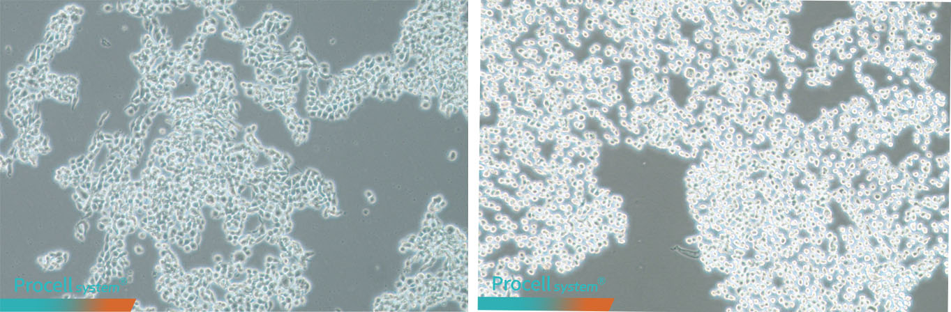
Figure 1. Representative cellular morphology at the optimal dissociation endpoint
(Left: LX-2; Right: A549)
IV. Cell Dissociation FAQ
Why do cells sometimes fail to detach completely?
Common causes include expired or improperly stored enzymes, repeated freeze-thaw cycles, low enzyme concentration, overconfluent cultures, and inadequate PBS washing, which leaves residual serum that inhibits enzyme activity.
Why can dissociation times vary between passages of the same cell line?
Protocol times are general references. Factors such as cell density, duration of adherent culture, enzyme brand, and storage conditions all affect dissociation. The endpoint should be determined by microscopic observation rather than strict timing.
Why do cell clumps appear after seeding?
Clumping is usually due to incomplete dissociation or insufficient pipetting. Causes include inadequate digestion, incomplete separation of intercellular junctions, insufficient resuspension after neutralization or centrifugation, or using an inappropriate pipette tip size. Large aggregates can impair attachment. Cells can be recollected, redigested, and gently resuspended before reseeding.
Can dissociation be performed at room temperature?
Yes. Trypsin is most active at 37℃, with slightly reduced activity at room temperature. Digestion time may need to be extended, and the endpoint should always be confirmed under a microscope to ensure complete dissociation.
Why do cell clumps become viscous and difficult to disperse?
Overdigestion can damage cells and release intracellular DNA, leading to aggregation and increased viscosity.
Adherent cell dissociation is not simply “add enzyme-wait-pipette”. Effective dissociation requires understanding cell adhesion biology, selecting appropriate reagents, and careful monitoring of the dissociation endpoint.
Next: Recommended Culture Medium Volumes and Cell Seeding Densities for Common Culture Vessels

