Key Details Not to Overlook in the Primary Isolation and Characterization of hUC-MSCs
Sep 24,2026
Key Details Not to Overlook in the Primary Isolation and Characterization of hUC-MSCs
In 2025, China's National Medical Products Administration (NMPA) granted conditional approval for Amimestrocel Injection, a human umbilical cord-derived mesenchymal stem cell product, for the treatment of a specific type of steroid-refractory acute graft-versus-host disease (aGVHD). This development has further increased interest in human umbilical cord mesenchymal stem cells (hUC-MSCs).
hUC-MSCs are primarily derived from Wharton's jelly of the umbilical cord. They exhibit strong proliferative capacity, multilineage differentiation potential, and relatively low immunogenicity. Compared with bone marrow- and adipose-derived sources, umbilical cord tissue is readily accessible and relatively abundant, and hUC-MSCs are now widely used in research on regenerative medicine, immunomodulation, and cell therapy.
However, obtaining stable, high-purity primary hUC-MSCs from a segment of umbilical cord involves much more than simply “cutting and culturing” the tissue. Donor screening, blood removal, vessel dissection, tissue explant placement, and early culture conditions can all directly affect subsequent cell attachment, proliferation, and phenotype. In this Cell Culture Academy feature, we use the tissue explant method as an example to walk through the key steps of primary hUC-MSC isolation.
Ⅰ. Sample Collection and Transport
Collect umbilical cord tissue from healthy full-term newborns in accordance with applicable ethical and biosafety requirements. To preserve cell viability, process the sample immediately after collection whenever possible. If transport is required, keep the tissue at a low temperature in a dedicated sterile preservation solution and avoid prolonged exposure to temperature fluctuations or environments with a high risk of contamination.
Ⅱ. Sample Preprocessing: The First Step in Primary Isolation Quality
1. Washing and Removing Residual Blood
Inside a biosafety cabinet, repeatedly wash the umbilical cord with sterile buffer (PBS + 1% penicillin-streptomycin) and gently compress it with forceps to remove residual blood from the vessels. If necessary, perform appropriate surface disinfection, such as soaking in 75% ethanol for 1 minute. After disinfection, rinse the tissue thoroughly with buffer several times to prevent residual disinfectant from damaging the cells.
Note: Residual red blood cells, platelets, and other blood components can contaminate Wharton's jelly, thereby affecting the viability and purity of the resulting cell population.
2. Removal of the Umbilical Arteries and Vein
Cut the umbilical cord into segments approximately 6 cm long. Make a longitudinal incision to expose two umbilical arteries and one umbilical vein. Carefully dissect the vessels using sterile instruments, keeping the vascular tissue as intact as possible while removing it completely (Figure 1).
Note: Vascular tissue contains smooth muscle cells, fibroblasts, and other cell types. Incomplete removal may result in contamination by non-target cells during subsequent culture. These cells may compete with hUC-MSCs for nutrients and may even inhibit hUC-MSC attachment and proliferation.
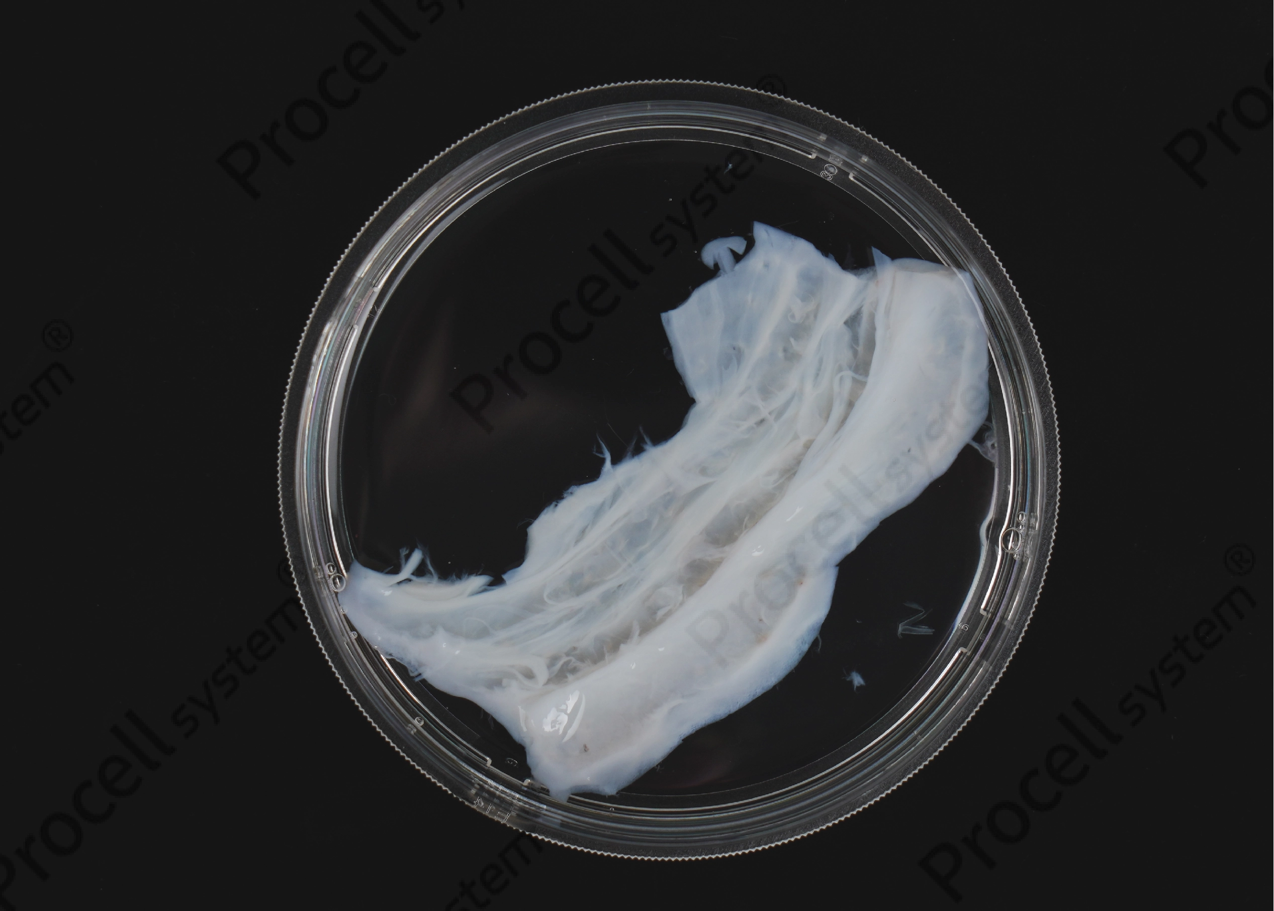
Figure 1. Umbilical Cord Tissue After Complete Removal of the Blood Vessels
3. Removal of the Amnion
Use sterile forceps to peel away the outer amniotic membrane while retaining the underlying Wharton's jelly. Note: This minimizes the introduction of non-target tissue and helps maintain the purity of the subsequently isolated hUC-MSCs.
4. Mincing the Wharton's Jelly
Cut the Wharton's jelly into relatively uniform tissue pieces, typically approximately 3–5 mm³, and thoroughly wash them with sterile buffer.
Note: Tissue pieces that are too large may hinder cell migration from the interior to the surface, whereas pieces that are too small or thin may be prone to floating during culture. Uniform, moderate-sized tissue pieces are therefore more suitable for subsequent explant culture.
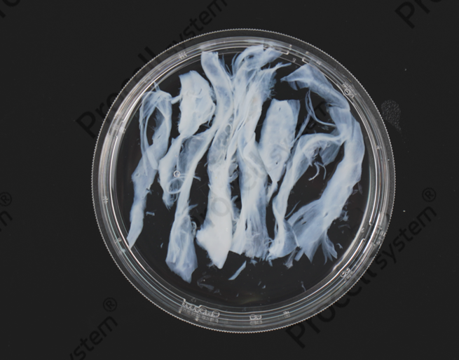
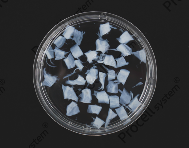
Figure 2. Wharton's Jelly Torn into Strips (Left) and Cut into Uniform Tissue Pieces (Right)
Ⅲ. Tissue Explant Culture and Passaging
Adherent culture: Evenly distribute the tissue pieces across the bottom of a culture flask, add complete medium suitable for hUC-MSC culture, and incubate at 37°C in a 5% CO₂ incubator.
The key to the tissue explant method is to provide a stable attachment environment that allows cells to gradually migrate from the edges of the tissue pieces and attach to the culture surface. Avoid moving the culture flask during the early culture period (the first 7 days) whenever possible. Once the tissue pieces are firmly attached, gently replace the medium.
As culture progresses, spindle-shaped or stellate cells can be observed migrating from the edges of the tissue pieces and forming parallel or whorled patterns. Continue culturing after medium replacement until cell density gradually increases.
Enzymatic passaging: When cells reach 75%–80% confluence, remove the remaining tissue pieces before enzymatic dissociation and passaging. When feasible, the recovered tissue pieces can be used for a second round of explant culture to improve sample utilization.
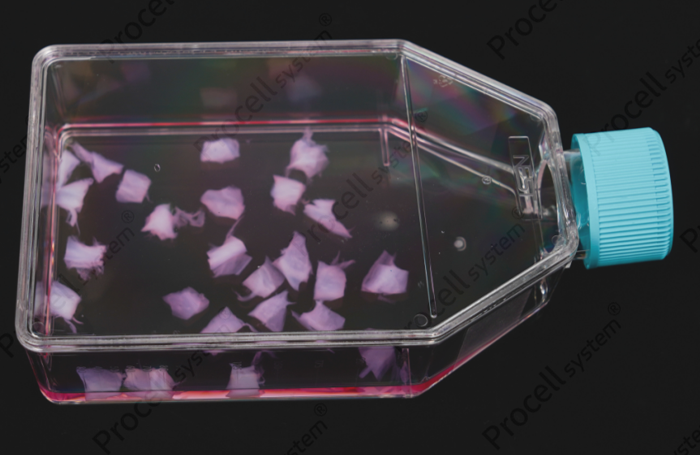
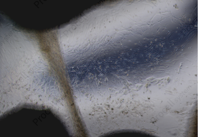
Figure 3. T75 Culture Flask with Inoculated Tissue Pieces (Left) and Microscopic Observation of Cells Migrating from the Tissue Pieces (Right)
Ⅳ. Key Considerations for Primary Isolation
1. Donor Screening: The Starting Point for Quality Control
Umbilical cord samples intended for research or subsequent development should undergo donor health screening according to project requirements, including assessment of infectious diseases and relevant genetic risks, to ensure that the sample source meets applicable ethical and quality standards.
2. Vessel Dissection: Slow and Careful Is Better Than Fast and Rough
The umbilical vein has a relatively thin wall. Avoid puncturing the vessel with the tips of the forceps and causing blood leakage during dissection, while removing the vascular tissue as completely as possible. Thorough vessel removal is an important step in minimizing contamination by non-target cells.
3. Gentle Handling: Minimize Mechanical Damage
Minimize unnecessary mechanical damage when dissecting the vessels, removing the amnion, and mincing the Wharton's jelly. Thoroughly remove residual blood to reduce its impact on subsequent culture.
4. Stable Explant Attachment: Critical During Early Culture
Use uniform tissue pieces approximately 3–5 mm³ in size, leave an appropriate space between pieces, and avoid moving the culture flask during the early culture period whenever possible. If no obvious cell outgrowth is observed after approximately 7 days of culture, investigate the possible causes based on tissue attachment, sample condition, and culture conditions, and re-plate the explants if necessary.
5. Culture System: Use Serum-Containing Media with Caution
Serum can promote cell differentiation. For experiments requiring maintenance of a specific cell phenotype or functional studies, the use of a culture-validated serum-free medium for mesenchymal stem cells is recommended.
6. Enzymatic Passaging: Both Enzyme Selection and Digestion Time Matter
Common enzymes used for hUC-MSC passaging include 0.25% trypsin (with EDTA), recombinant trypsin, and Accutase Cell Detachment Solution. If downstream assays involve cell phenotype or surface antigen analysis, the relatively gentle Accutase Cell Detachment Solution or recombinant trypsin may be preferred. For routine expansion, conventional trypsin can also be used. Regardless of the dissociation reagent selected, the degree of digestion should be carefully controlled to avoid excessive digestion, which may cause cell damage and alter surface antigen expression.
Ⅴ. Quality Control and Cell Characterization
After obtaining the cells, multiple parameters should be evaluated to determine whether they meet the criteria for hUC-MSCs.
1. Morphological Assessment
Under microscopic observation, the cells typically exhibit a fibroblast-like, spindle-shaped, or fusiform morphology and may form parallel or whorled patterns.
2. Flow Cytometric Characterization of Surface Markers
Typical hUC-MSCs should highly express mesenchymal stem cell-positive markers such as CD73, CD90, and CD105 (positive rate ≥95%), while showing low or no expression of hematopoietic stem cell and lineage markers such as CD34 and CD45 (positive rate ≤2%).
3. Multilineage Differentiation Potential
Under specific induction conditions, the cells should be capable of differentiating into osteoblasts, chondrocytes, and adipocytes.
Primary isolation of hUC-MSCs is not simply a matter of tissue processing. From sample screening and tissue preprocessing to explant culture and passaging, every step can affect the final cell quality. After cell isolation, characterization should also incorporate morphology, surface markers, and multilineage differentiation potential. Standardized procedures and comprehensive quality control are the foundation for obtaining stable, high-quality hUC-MSCs and provide a solid basis for subsequent research and applications.
Prev: Exosome Purification and Characterization: Key Considerations for High-Quality Samples